Course Code: BP104T
Unit III Introduction
Body fluids, blood and lymphatic system; inflammation and repair; haematological diseases. This unit explains how the body transports substances, produces blood cells, limits bleeding, recovers tissue fluid and responds to injury. Normal physiology provides the basis for understanding the required blood disorders.
Blood is a fluid connective tissue. Its plasma transports dissolved substances, while formed elements carry respiratory gases, defend tissues and support haemostasis. Lymphatic channels return interstitial fluid and proteins to the circulation and connect tissues to immune organs.
Inflammation recruits defensive cells and mediators to damaged tissue. An effective response removes the cause and permits repair; excessive or persistent inflammation itself causes injury.
Topics Covered – Complete Syllabus Map
| Required topic | Sections |
|---|---|
| Body-fluid compartments | 3.1 |
| Composition and functions of blood | 3.2–3.5 |
| Haemopoiesis | 3.6–3.7 |
| Formation of haemoglobin | 3.8 |
| Coagulation mechanisms | 3.9–3.11 |
| Blood grouping, Rh and transfusion | 3.12–3.13 |
| Lymphatic organs, tissues and vessels | 3.14–3.17 |
| Lymph formation, circulation and functions | 3.15, 3.18–3.19 |
| Inflammation: introduction and classification | 3.20 |
| Pathophysiology and mediators | 3.21–3.24 |
| Iron-deficiency and megaloblastic anaemia | 3.25–3.27 |
| Sickle-cell anaemia and thalassaemia | 3.28–3.29 |
| Hereditary/acquired anaemia; haemophilia | 3.30–3.31 |
| Cases, revision and exam practice | 3.32–3.33 |
Learning Outcomes
- Locate intracellular, interstitial and plasma compartments.
- Identify blood cells by structure and explain their functions.
- Trace blood-cell production and haemoglobin synthesis.
- Explain platelet-plug formation, coagulation and fibrinolysis.
- Interpret ABO and Rh grouping and distinguish component compatibility.
- Draw lymphatic vessels, a lymph node and the spleen with meaningful labels.
- Explain vascular and cellular events in inflammation.
- Compare the mechanisms of the specified anaemias and haemophilia.
Essential Vocabulary
| Term | Meaning |
|---|---|
| Haematocrit | Fraction of blood volume occupied by RBCs |
| Haemopoiesis | Production of blood’s formed elements |
| Erythropoietin | Hormone promoting erythroid production |
| Haemostasis | Processes limiting blood loss |
| Agglutination | Antibody-mediated clumping of cells |
| Lymph | Interstitial fluid inside lymphatic vessels |
| Exudate | Inflammatory, protein-rich extravascular fluid |
| Diapedesis | Leukocyte passage across vessel wall |
| Anaemia | Reduced haemoglobin concentration |
| Haemolysis | Premature destruction of RBCs |
Study Method
Normal structure → normal function → disturbed mechanism → consequence
For each disease, connect the defect to the blood-film appearance and the clinical effects. Learn compatibility tables for the stated component rather than applying one table to every transfusion product.
Central question: What happens when oxygen carriage, coagulation, tissue-fluid drainage or the inflammatory response fails?
3.1 Body Fluids and Compartments
Total body water is distributed inside and outside cells. In a typical lean adult male, water is approximately 60% of body mass; age, sex and body-fat proportion alter this value. Infants generally have a higher fraction and older adults a lower one. These are teaching estimates, not universal constants.
Main Compartments
| Compartment | Approximate share of total body water | Main location |
|---|---|---|
| Intracellular fluid, ICF | Two-thirds | Within cells |
| Extracellular fluid, ECF | One-third | Outside cells |
| Interstitial fluid | About three-quarters of ECF | Between tissue cells |
| Plasma | About one-quarter of ECF | Within blood vessels |
Small specialised spaces contain transcellular fluid, including cerebrospinal, synovial and serous fluids. Depending on the teaching model, these are treated separately within ECF.
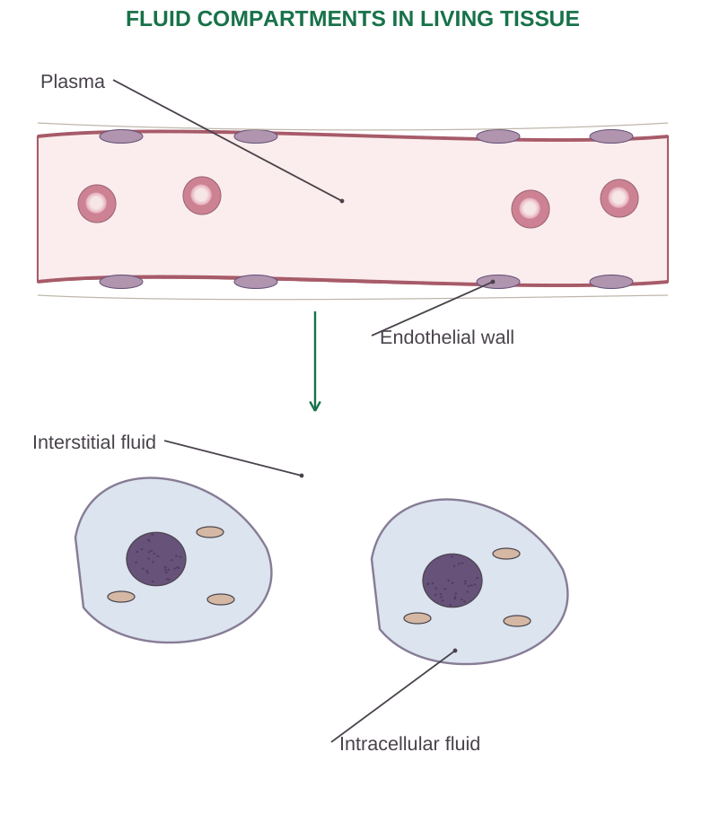
Diagram labels: Plasma; endothelial wall; interstitial fluid; intracellular fluid.
Movement and Composition
Cell membranes separate ICF from ECF; capillary walls separate plasma from tissue fluid. Water crosses many membranes by osmosis, whereas solute movement depends on permeability, transporters and gradients.
ICF is relatively rich in potassium and intracellular phosphates; ECF is relatively rich in sodium and chloride. Plasma has more protein than ordinary interstitial fluid.
Worked Example
For a 70 kg adult using a 60% water estimate:
- Total water ≈ 42 L.
- ICF ≈ 28 L.
- ECF ≈ 14 L.
- Interstitial fluid ≈ 10.5 L.
- Plasma ≈ 3.5 L.
These values explain proportions; actual measurements vary.
Fluid Balance and Oedema
Intake and metabolic production balance losses through kidneys, skin, lungs and intestine. Oedema is excess interstitial fluid. Raised hydrostatic pressure, reduced plasma oncotic pressure, increased vascular permeability or impaired lymphatic drainage can produce it.
Albumin helps retain water within plasma, but fluid exchange also depends on the endothelial barrier and its glycocalyx.
Avoid a common shortcut: Do not say that every filtered drop returns at the venous end of each capillary. Sustained tissue-fluid recovery depends importantly on lymphatic drainage.
3.2 Blood: Composition and Physical Features
Blood comprises plasma and formed elements. Plasma is the fluid extracellular matrix. Formed elements are erythrocytes, leukocytes and platelets; platelets are cell fragments rather than complete nucleated cells.
| Feature | Typical adult teaching value |
|---|---|
| Blood volume | Roughly 5 L; depends on body size |
| pH | Approximately 7.35–7.45 |
| Temperature | Slightly above average body temperature |
| Plasma fraction | Approximately 55% in an illustrative sample |
| Formed-element fraction | Approximately 45%, mainly RBCs |
Haematocrit
The haematocrit is the volume fraction of RBCs in blood. After centrifugation of anticoagulated blood, RBCs settle below a thin buffy coat of leukocytes and platelets, with plasma above. The buffy coat is normally less than 1% of the sample volume.
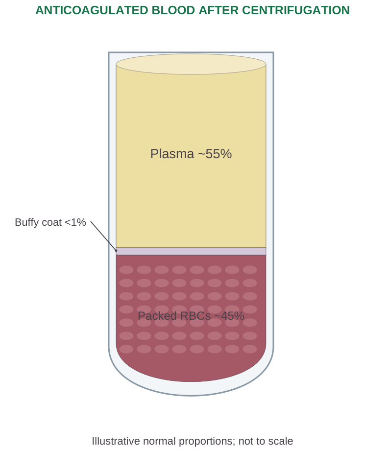
Diagram labels: Plasma ~55%; buffy coat <1%; packed RBCs ~45%. Illustrative normal proportions, not to scale.
Haematocrit (%) = packed RBC volume ÷ total blood volume × 100
If 4.2 mL of a 10 mL sample is packed RBCs, haematocrit = 42%. The remaining volume includes both plasma and buffy coat.
Physiological Relevance
More RBCs generally raise blood viscosity. Dehydration can increase measured haemoglobin and haematocrit by reducing plasma volume, without creating extra RBCs. Conversely, plasma-volume expansion can lower measured concentrations. Interpret results with hydration and clinical context.
3.3 Plasma Constituents and Blood Functions
Plasma is mainly water, with proteins, electrolytes, nutrients, metabolic waste, dissolved gases and signalling molecules.
| Component | Main importance |
|---|---|
| Albumin | Major contributor to plasma oncotic pressure; carrier protein |
| Globulins | Transport functions; immunoglobulins provide antibody defence |
| Fibrinogen | Soluble precursor of fibrin in coagulation |
| Electrolytes | Osmotic balance, excitability and acid–base regulation |
| Nutrients and hormones | Transport to target tissues |
| Urea and other waste | Carriage to excretory organs |
Plasma Versus Serum
Plasma is obtained when clotting is prevented.
Serum is the liquid remaining after blood has clotted; fibrinogen and some coagulation components have been consumed or removed with the clot. Serum is not simply plasma with all proteins removed.
Functions of Blood
- Transport: Oxygen, carbon dioxide, nutrients, hormones and waste.
- Regulation: Acid–base balance, temperature distribution and fluid exchange.
- Protection: Leukocyte and antibody defence, platelet plugs and clot formation.
The components cooperate: plasma carries cells and substrates; RBCs carry most oxygen; WBCs defend tissues; platelets and clotting proteins limit bleeding.
3.4 Erythrocytes: Structure and Functions
Mature human RBCs are biconcave, anucleate discs, about 7–8 µm across. Their thin central region produces central pallor in a stained blood film. They lack mitochondria and obtain ATP mainly by glycolysis, helping them avoid consuming the oxygen they transport.
Why Their Structure Matters
A high surface-area-to-volume ratio assists gas exchange. A flexible cytoskeleton allows deformation in narrow capillaries. Haemoglobin binds oxygen reversibly; RBC enzymes and haemoglobin also contribute to carbon-dioxide carriage and buffering.
Life Cycle
Average RBC lifespan is about 120 days. Macrophages, especially in spleen, liver and marrow, remove ageing cells.
Iron is recycled, globin is broken into amino acids, and haem is converted through biliverdin to bilirubin. Unconjugated bilirubin travels bound to albumin before liver processing and biliary excretion.
Oxygen Carriage
Most oxygen is haemoglobin-bound; a small amount dissolves in plasma. Oxygen saturation describes the fraction of available haemoglobin binding sites occupied by oxygen.
Normal saturation does not exclude anaemia: too little haemoglobin can limit oxygen content even when its remaining sites are well saturated.
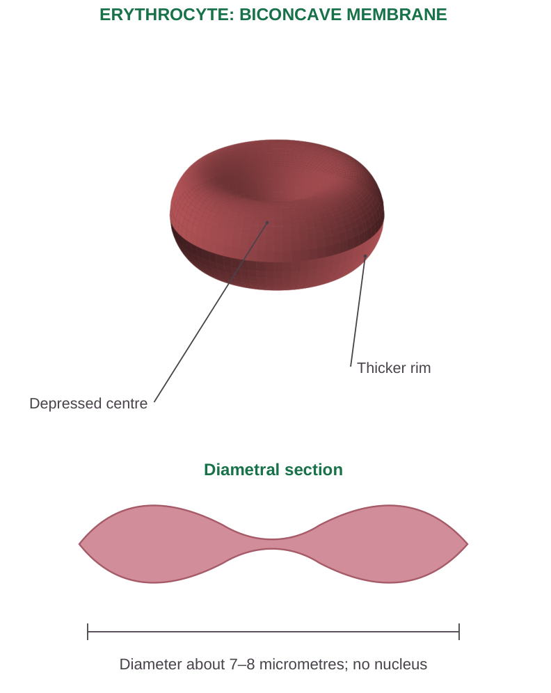
Diagram labels: Depressed centre; thicker rim; diametral section. Diameter about 7–8 micrometres; no nucleus.
3.5 Leukocytes and Platelets
Leukocytes possess nuclei and participate in host defence. Granulocytes have conspicuous specific granules; agranulocytes lack those conspicuous granules, although they still contain other cellular granules.
| Cell | Identifying feature | Main role |
|---|---|---|
| Neutrophil | Multilobed nucleus, fine granules | Rapid phagocytic response, especially to bacteria |
| Eosinophil | Usually bilobed nucleus, coarse eosinophilic granules | Parasite defence and allergic inflammation |
| Basophil | Dark granules can obscure nucleus | Mediator release in allergic responses |
| Lymphocyte | Dense round nucleus with variable cytoplasm | B-, T- and NK-cell immune functions |
| Monocyte | Large cell, folded/kidney-shaped nucleus | Tissue macrophage and related populations |
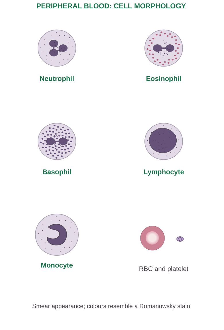
Diagram labels: Neutrophil; eosinophil; basophil; lymphocyte; monocyte; RBC and platelet. Smear appearance; colours resemble a Romanowsky stain.
Platelet Structure and Role
Platelets are small anucleate fragments released from marrow megakaryocytes. Their cytoskeleton changes shape during activation; granules release mediators, and surface phospholipids support coagulation reactions. Their circulating lifespan is approximately 7–10 days.
A teaching platelet count is about 150–450 × 10^9/L, but the reporting laboratory supplies its own interval.
Low count or impaired function often causes petechiae and mucosal bleeding. Coagulation-factor defects more often cause deep-tissue bleeding; this distinction is useful but not absolute.
3.6 Haemopoiesis: Origin and Sites
Haemopoiesis is production of the formed elements of blood from haematopoietic stem cells. Stem cells self-renew and produce progressively restricted progenitors. Differentiation requires a supportive marrow microenvironment, growth factors and adequate nutrients.
Sites Through Life
Early embryonic blood formation begins in the yolk sac. The fetal liver becomes a major site, with other fetal tissues contributing; bone marrow progressively takes over.
Adult production is mainly in red marrow of the pelvis, vertebrae, sternum, ribs, skull and proximal regions of selected long bones.
Major Lineages
| Lineage | Principal mature products |
|---|---|
| Myeloid | RBCs; megakaryocytes/platelets; granulocytes; monocytes |
| Lymphoid | B cells, T cells and NK cells |
This division is a useful introductory model; lineage decisions in living marrow are more complex than a rigid two-branch chart. B-cell development is associated with marrow; T-cell maturation requires the thymus.
Regulation
Erythropoietin supports erythroid progenitors. Thrombopoietin, produced mainly by the liver, supports megakaryocytes and platelets. Colony-stimulating factors and interleukins support several leukocyte populations.
Extramedullary haemopoiesis means blood-cell formation outside marrow, for example in liver or spleen in some severe disorders.
3.7 Erythropoiesis and Feedback
Erythropoiesis produces RBCs. As erythroid precursors mature, cells become smaller, chromatin condenses, haemoglobin accumulates and the nucleus is expelled. A reticulocyte retains some RNA before becoming a mature erythrocyte.
Recognisable Sequence
Proerythroblast → basophilic erythroblast → polychromatophilic erythroblast → orthochromatic erythroblast → reticulocyte → mature RBC
Early precursors divide; late maturation includes nuclear extrusion rather than repeated division of mature RBCs.
Oxygen-Sensing Feedback
- Step 1: Reduced renal oxygen availability is sensed.
- Step 2: Renal interstitial cells increase erythropoietin output.
- Step 3: EPO stimulates survival and production of erythroid precursors.
- Step 4: More circulating RBCs improve oxygen delivery, reducing the stimulus.
Requirements and Interpretation
Iron supplies haem synthesis; amino acids supply globin; vitamin B12 and folate support DNA synthesis.
The reticulocyte response helps distinguish appropriate marrow compensation from impaired production. Renal disease can lower EPO, whereas haemolysis often raises reticulocytes if marrow function and nutrients remain adequate.
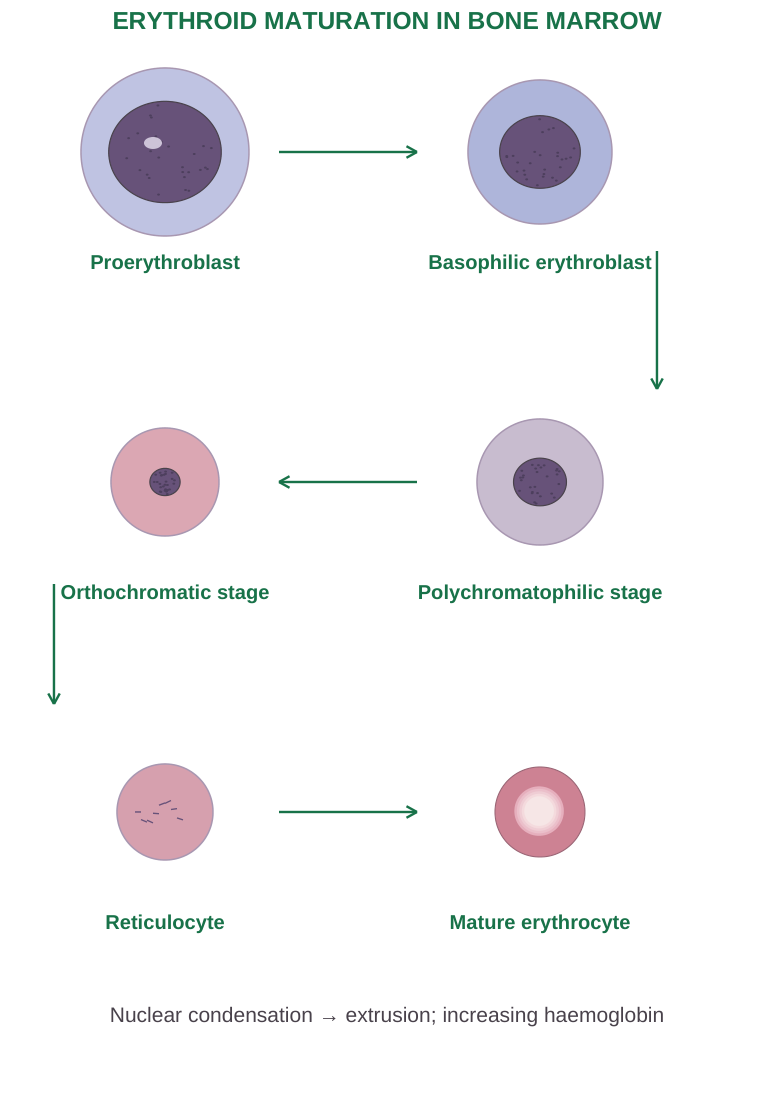
Diagram labels: Proerythroblast; basophilic erythroblast; polychromatophilic stage; orthochromatic stage; reticulocyte; mature erythrocyte. Nuclear condensation → extrusion; increasing haemoglobin.
3.8 Formation and Functions of Haemoglobin
Haemoglobin is a tetramer of four globin chains, each associated with a haem group. Each haem contains ferrous iron, Fe2+, capable of reversibly binding one oxygen molecule; one haemoglobin molecule can therefore bind up to four O2 molecules.
Synthesis in Erythroid Precursors
Haem formation uses both mitochondria and cytosol. Glycine and succinyl-CoA form aminolevulinic acid through ALA synthase. Subsequent reactions produce porphyrin intermediates and protoporphyrin IX. Ferrochelatase inserts Fe2+ to form haem. Globin chains are synthesised on ribosomes and assemble with haem.
Mature RBCs lack the organelles needed for new haemoglobin synthesis. Most synthesis occurs before full erythrocyte maturation; reticulocytes retain limited residual synthetic capacity during their final maturation.
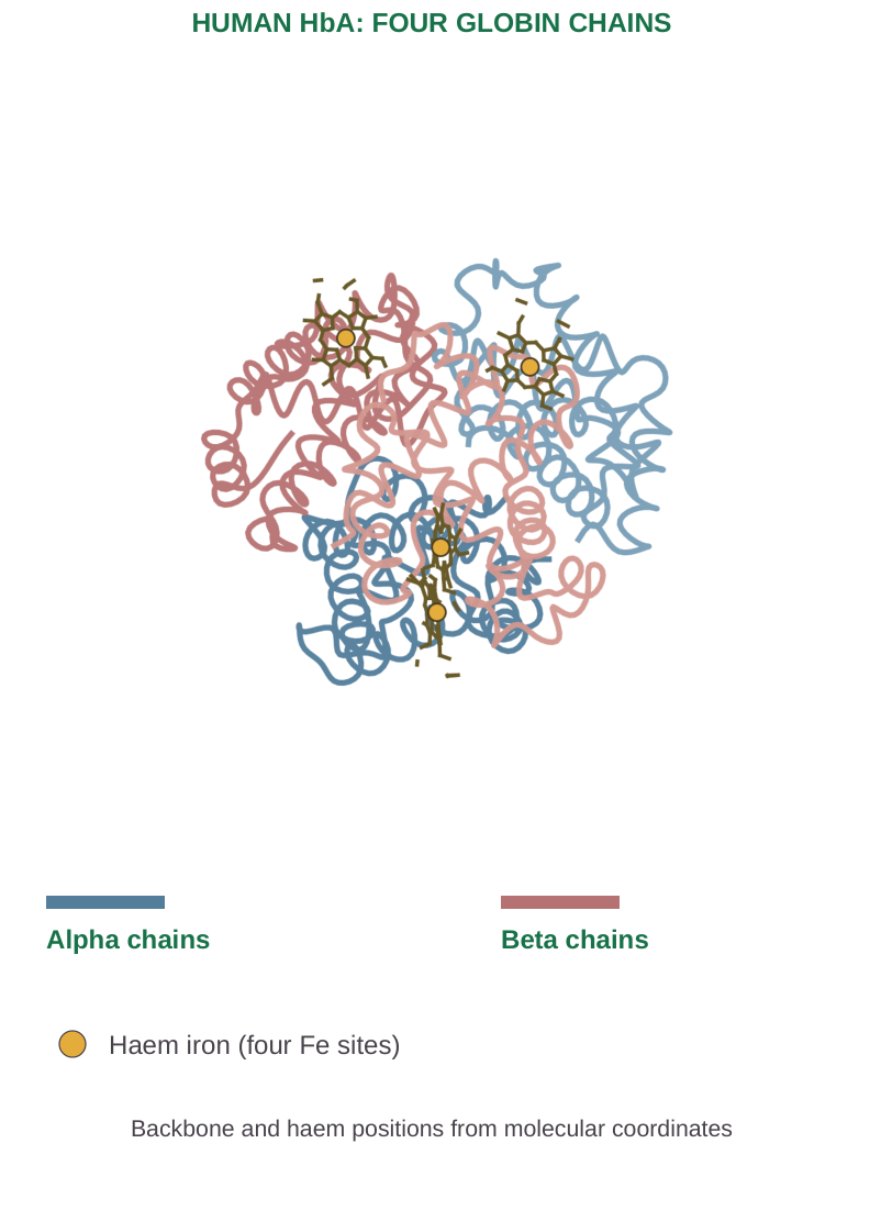
Diagram labels: Human HbA: four globin chains. Alpha chains; beta chains; haem iron, four Fe sites. Backbone and haem positions from molecular coordinates.
| Haemoglobin | Globin composition | Teaching significance |
|---|---|---|
| HbA | α2β2 | Predominant adult haemoglobin |
| HbA2 | α2δ2 | Minor adult component |
| HbF | α2γ2 | Predominant fetal form; higher O2 affinity |
Functional Behaviour
Oxygen binding is cooperative: binding at one site changes the affinity of other sites. The oxygen-dissociation curve is sigmoid. Increased CO2, acidity and temperature favour oxygen unloading in active tissues. HbF’s greater affinity helps fetal uptake from maternal blood.
Disorders Connected to Synthesis
Iron shortage reduces haem availability. Thalassaemia reduces synthesis of a globin-chain type. Sickle-cell disease changes β-globin structure. These different molecular problems can all cause anaemia, but they do not share the same mechanism.
Exam distinction: Oxygenation is reversible O2 binding to ferrous haem. Oxidation of iron to Fe3+ produces methaemoglobin, which cannot carry oxygen normally.
3.9 Haemostasis: Response to Vascular Injury
Haemostasis limits blood loss at an injured vessel. It includes local vasoconstriction, platelet-plug formation and fibrin formation, followed by clot retraction and eventual removal. Primary haemostasis and coagulation overlap rather than occurring as completely isolated events.
Vascular Spasm
Vessel injury triggers local contraction through neural, muscular and chemical influences. Reduced flow helps the developing plug remain at the damaged site. This response alone is insufficient for many injuries.
Primary Haemostasis: Platelet Plug
- Step 1: Injury exposes collagen and associated subendothelial material.
- Step 2: von Willebrand factor supports platelet adhesion, particularly through platelet GPIb.
- Step 3: Platelets activate, change shape and release granule mediators; ADP and thromboxane A2 recruit further platelets.
- Step 4: Activated GPIIb/IIIa binds fibrinogen, bridging platelets during aggregation.
Secondary Haemostasis
Coagulation generates thrombin and then fibrin, reinforcing the temporary platelet plug. A platelet-rich plug without adequate fibrin is mechanically less stable. Fibrin is a protein mesh, not a solid lid placed over the vessel.
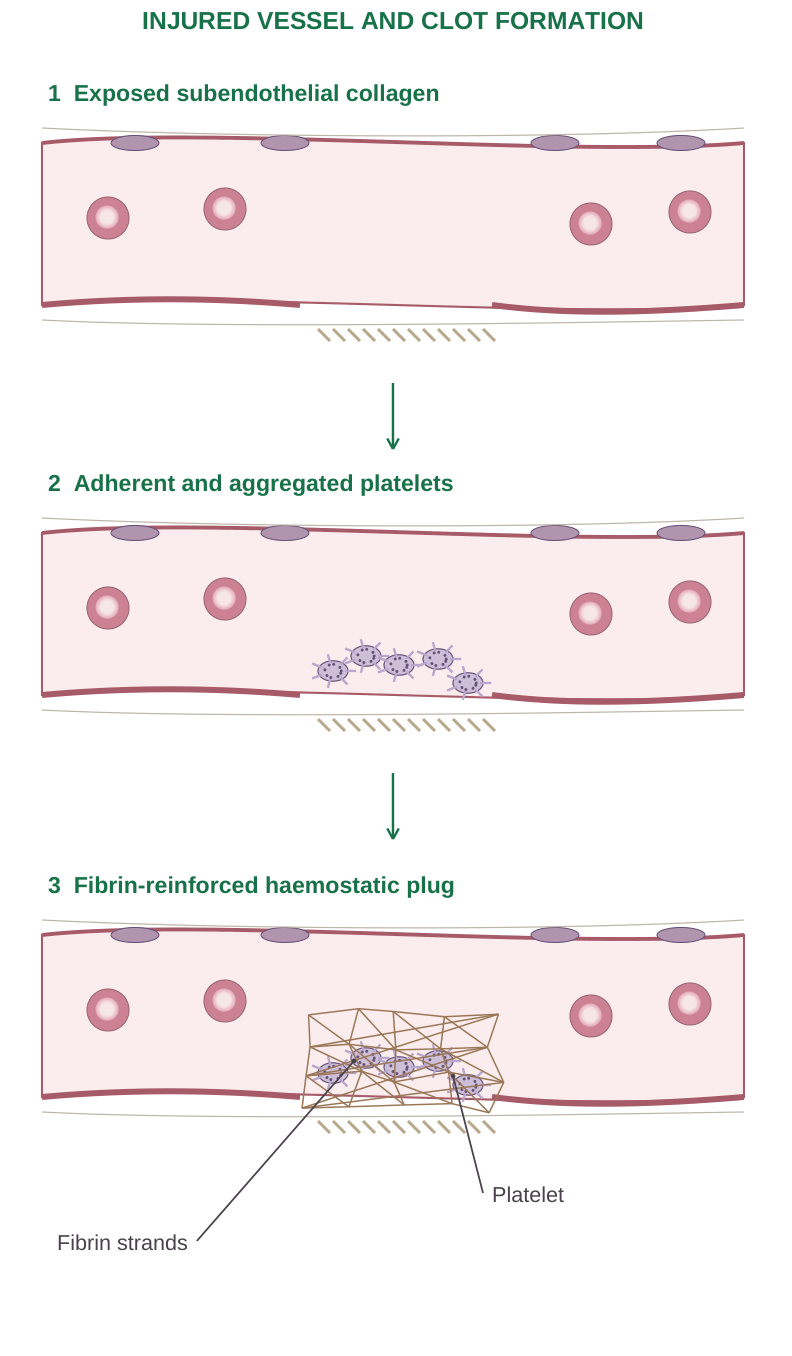
Diagram labels: Exposed subendothelial collagen; adherent and aggregated platelets; fibrin-reinforced haemostatic plug; fibrin strands; platelet.
3.10 Coagulation: Pathways and Common Mechanism
The classical cascade separates extrinsic, intrinsic and common pathways. It is valuable for learning factor relationships and laboratory tests. In vivo, tissue-factor initiation, platelet surfaces and amplification cooperate; the pathways are not independent pipes carrying blood.
| Teaching pathway | Main sequence |
|---|---|
| Extrinsic | Tissue factor with VIIa activates X; can also activate IX |
| Intrinsic | XII → XI → IX; IXa with VIIIa activates X |
| Common | Xa with Va converts II to IIa; thrombin forms fibrin from I; XIIIa cross-links fibrin |
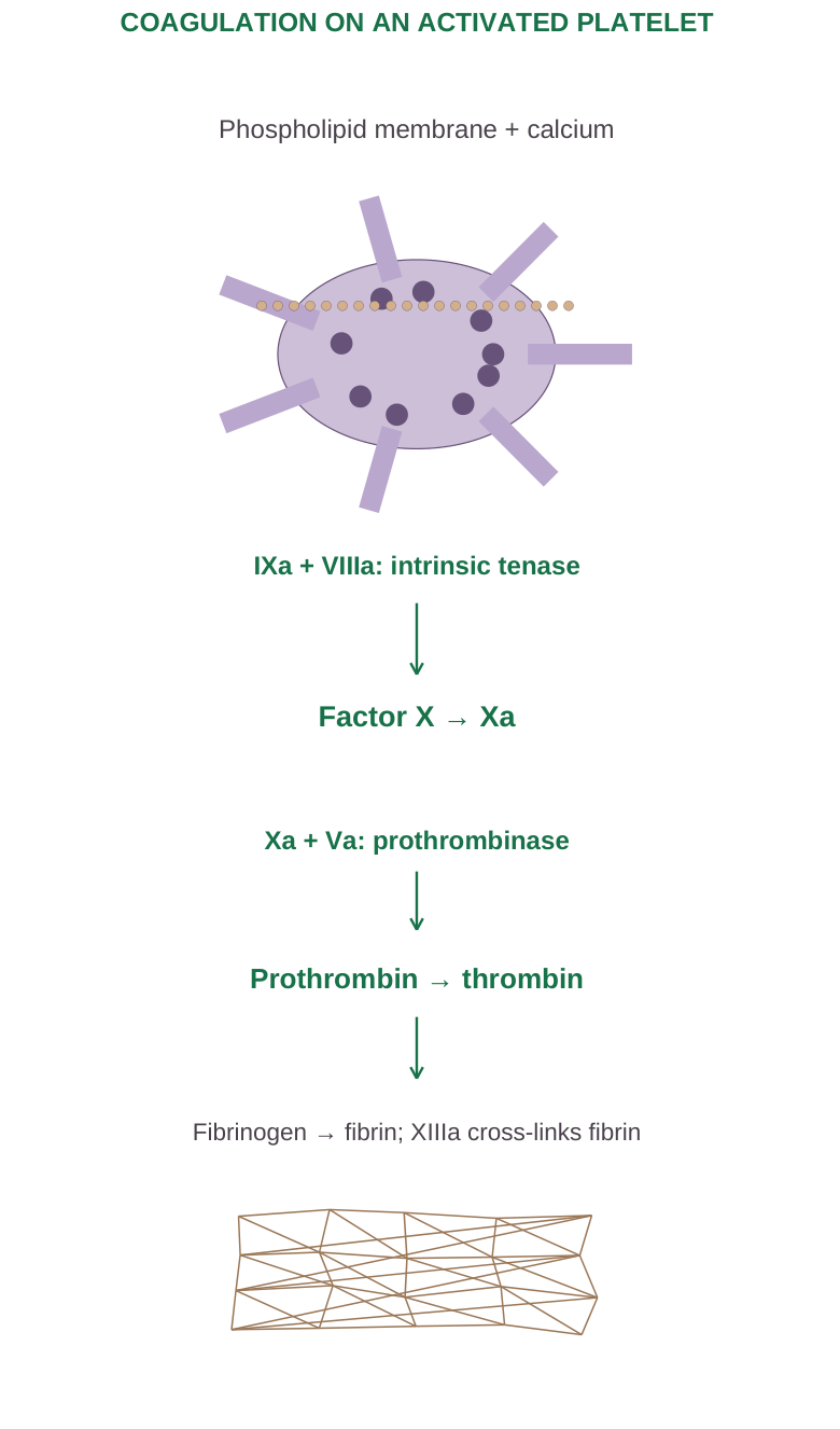
Diagram labels: Phospholipid membrane + calcium; IXa + VIIIa: intrinsic tenase; factor X → Xa; Xa + Va: prothrombinase; prothrombin → thrombin; fibrinogen → fibrin; XIIIa cross-links fibrin.
Common Final Reactions
Prothrombin (II) → thrombin (IIa).
The prothrombinase complex includes Xa, Va, calcium and a suitable phospholipid surface.
Fibrinogen (I) → fibrin.
Thrombin cleaves fibrinogen, allowing fibrin assembly; activated factor XIII makes cross-links that stabilise the mesh.
Thrombin also activates platelets and several factors, amplifying the response. Calcium and membrane surfaces organise reactions locally. Factor XII is important in the laboratory contact pathway; its deficiency does not usually cause abnormal bleeding, illustrating the limits of the simplified cascade.
Vitamin K and Laboratory Tests
Vitamin K supports functional maturation of factors II, VII, IX and X, and proteins C and S.
PT primarily assesses the extrinsic/common teaching pathways; aPTT assesses intrinsic/common pathways. The INR standardises PT for selected uses. A prolonged test is a clue, not a complete diagnosis.
3.11 Clot Control, Retraction and Fibrinolysis
Haemostasis must remain local. Intact endothelium discourages platelet activation and excessive coagulation through surface properties and mediators such as nitric oxide and prostacyclin.
Physiological Anticoagulant Mechanisms
- Antithrombin inhibits thrombin and several activated coagulation factors.
- Protein C with protein S inactivates Va and VIIIa after appropriate endothelial activation.
- Tissue-factor pathway inhibitor limits tissue-factor-associated initiation.
- Blood flow dilutes and removes activated mediators from the injury site.
Clot Retraction
Platelet contractile proteins pull on the fibrin network, drawing the wound margins closer and expelling fluid from the clot. Platelets therefore contribute both to early aggregation and later mechanical consolidation.
Fibrinolysis
Tissue plasminogen activator helps convert plasminogen to plasmin on fibrin. Plasmin breaks the fibrin network into degradation products. Inhibitors regulate activators and plasmin so that clot removal is controlled.
D-dimer is produced when cross-linked fibrin is degraded; an increased result is not specific to one disease.
Important Distinctions
A thrombus forms within a vessel or heart; an embolus is material carried through blood to another site.
Physiological haemostasis is local and protective, whereas inappropriate thrombosis can obstruct perfusion. Excess bleeding and excessive clotting represent different failures of the same regulated system.
3.12 ABO Blood Grouping
Blood groups reflect antigens on RBC membranes. In the ABO system, naturally occurring antibodies develop against the absent major ABO antigen, influenced by environmental exposure. ABO antibodies are mainly IgM; clinically important exceptions exist, particularly some IgG in group O individuals.
| ABO group | RBC antigens | Usual plasma antibodies |
|---|---|---|
| A | A | Anti-B |
| B | B | Anti-A |
| AB | A and B | Neither anti-A nor anti-B |
| O | Neither A nor B | Anti-A and anti-B |
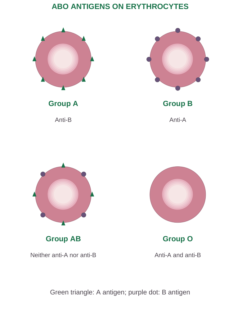
Diagram labels: Group A: anti-B. Group B: anti-A. Group AB: neither anti-A nor anti-B. Group O: anti-A and anti-B. Green triangle: A antigen; purple dot: B antigen.
Agglutination and Grouping
Mixing RBCs with reagent anti-A or anti-B demonstrates the corresponding antigen when clumping occurs. Forward typing tests RBC antigens; reverse typing tests expected plasma antibodies. Results must agree appropriately before the group is accepted.
Packed-RBC Compatibility: ABO Only
| Recipient | ABO RBC groups that can be compatible |
|---|---|
| O | O |
| A | A or O |
| B | B or O |
| AB | AB, A, B or O |
These are introductory ABO rules for red-cell components, not permission to bypass Rh assessment, antibody screening or crossmatching.
Scientist Connection
Karl Landsteiner’s blood-group discoveries established why apparently similar human blood samples could be incompatible. His work provided a scientific basis for safer transfusion; he received the 1930 Nobel Prize in Physiology or Medicine.
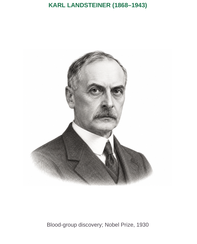
Diagram labels: Karl Landsteiner, 1868–1943. Blood-group discovery; Nobel Prize, 1930.
3.13 Rh Factors and Transfusion Principles
Rh is a blood-group system with several antigens. Routine Rh-positive/Rh-negative terminology usually refers to the D antigen. A D-negative person generally does not possess natural anti-D; exposure to D-positive RBCs can produce immune anti-D.
Maternal–Fetal Incompatibility
Maternal sensitisation can follow exposure to fetal RBCs or previous transfusion. Maternal IgG anti-D can cross the placenta and destroy fetal D-positive RBCs, producing haemolytic disease of the fetus and newborn.
Risk depends on sensitisation and fetal antigen status, not merely on different maternal and paternal blood groups.
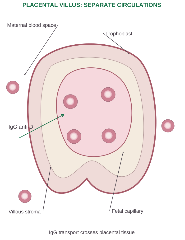
Diagram labels: Maternal blood space; trophoblast; villous stroma; fetal capillary; IgG anti-D. IgG transport crosses placental tissue.
Anti-D immunoglobulin is used in eligible unsensitised D-negative pregnancies to reduce sensitisation. It prevents immune development; it does not treat established maternal alloimmunisation by removing existing anti-D.
Component-Specific Compatibility
For plasma, the major ABO concern is donor antibodies acting on recipient RBCs. AB plasma lacks anti-A and anti-B, so the simplified plasma-donor rule reverses the RBC rule.
Whole blood, platelets and other components require their own transfusion-service policies; an RBC table cannot be applied to all of them.
Safe-Transfusion Workflow
- Step 1: Establish identity, indication and required component.
- Step 2: Determine ABO/Rh type, screen relevant antibodies and perform required compatibility testing.
- Step 3: Recheck patient and component identifiers before administration.
- Step 4: Monitor according to protocol for acute reactions.
Acute ABO incompatibility may cause intravascular haemolysis, fever, hypotension, renal injury and coagulation disturbance. A suspected reaction requires stopping the transfusion and immediate assessment under clinical protocol.
The phrase “universal donor” is a component-specific shorthand, not a guarantee of universal safety.
3.14 Lymphatic System: Components and Organisation
The lymphatic system comprises lymph, lymphatic capillaries and collecting vessels, trunks and ducts, lymphoid organs and lymphoid tissues. It returns escaped fluid and proteins to blood and supports immune surveillance.
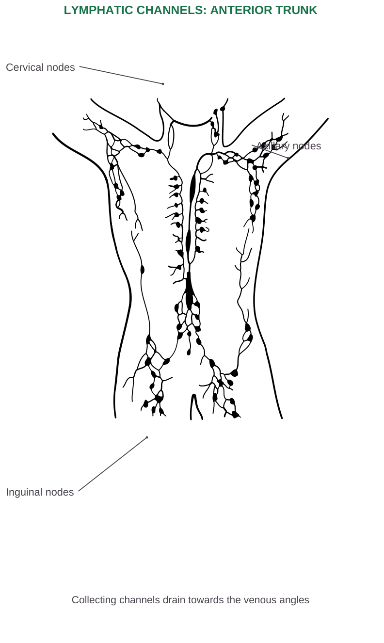
Diagram labels: Cervical nodes; axillary nodes; inguinal nodes. Collecting channels drain towards the venous angles.
Primary and Secondary Lymphoid Organs
Primary organs support lymphocyte development:
- Bone marrow for B-cell development.
- Thymus for T-cell maturation.
Secondary organs and tissues support encounters between mature lymphocytes and antigens: lymph nodes, spleen and mucosa-associated lymphoid tissue, including tonsils and intestinal lymphoid tissue.
Why It Is a Return System
Blood forms a closed circulatory circuit powered by the heart. Lymph begins in tissue spaces and follows a one-way return route toward veins.
Lymph nodes are situated along collecting vessels; the spleen filters blood rather than lying along the lymph-flow route.
3.15 Lymph Formation and Lymphatic Capillaries
Fluid that leaves blood microvessels enters tissue spaces. Interstitial fluid becomes lymph only after entering a lymphatic vessel.
Its composition reflects the drained tissue; intestinal lymph after fat absorption can be rich in chylomicrons and appear milky, termed chyle.
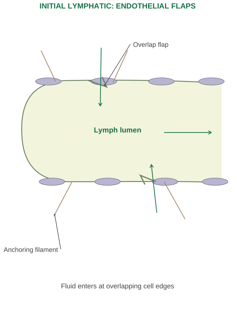
Diagram labels: Lymph lumen; anchoring filament; overlap flap. Fluid enters at overlapping cell edges.
Initial Lymphatic Structure
Initial lymphatics are blind-ended endothelial channels. Overlapping endothelial edges form flap-like entry regions. Anchoring filaments connect the vessel wall to surrounding connective tissue and help prevent collapse when interstitial pressure rises.
Entry and Flow
Raised interstitial pressure favours inward opening of endothelial flaps. Rising pressure inside the lymphatic discourages reverse entry flow.
Larger collecting vessels have smooth muscle and internal valves. Tissue movement and intrinsic vessel contraction help propel lymph through successive segments.
A Physiological Link
Lymphatics recover fluid, macromolecules, antigens and migrating immune cells. Obstruction reduces drainage and can produce lymphoedema.
Most brain tissue does not contain conventional lymphatic capillaries, although specialised CNS fluid drainage and meningeal lymphatic pathways exist.
3.16 Lymph Node: Structure and Lymph Filtration
A lymph node is an encapsulated, often bean-shaped organ. Afferent lymphatic vessels enter the convex surface; efferent lymphatics leave at the hilum.
Blood vessels also enter or leave at the hilum, but blood and lymph occupy separate pathways.
| Region | Main structure/function |
|---|---|
| Capsule and trabeculae | Connective-tissue support |
| Subcapsular sinus | Receives incoming lymph |
| Outer cortex | Lymphoid follicles, predominantly B-cell regions |
| Paracortex | Predominantly T-cell region; high endothelial venules |
| Medulla | Medullary cords and sinuses; plasma cells and macrophages |
| Hilum | Exit for efferent lymph; blood-vessel access |
Flow Through a Node
Afferent vessels → subcapsular sinus → cortical/trabecular sinuses → medullary sinuses → efferent vessel
Reticular support and resident cells facilitate antigen sampling and filtration rather than acting as an inert sieve alone.
Immune Role
Antigen-presenting cells interact with lymphocytes; activated populations proliferate and may form germinal centres. Lymphocytes can enter nodes from blood through high endothelial venules.
Enlarged nodes may indicate immune activation, infection or other disorders; enlargement alone does not identify the cause.
Labels in the lymph-node illustration: Afferent lymph vessels; trabeculae; medullary sinus; cortical sinus; medulla; capsule; subcapsular sinus; blood vessel entering the hilum; slow flowing lymph; lymphocytes in outflowing lymph; lymphocyte; efferent lymph vessel; reticular fiber.
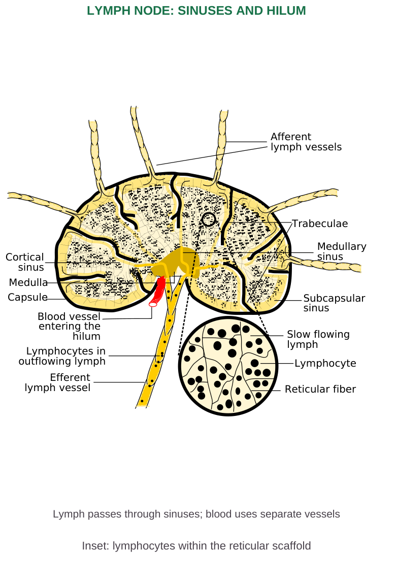
Diagram labels: Lymph node: sinuses and hilum. Lymph passes through sinuses; blood uses separate vessels. Inset: lymphocytes within the reticular scaffold.
3.17 Spleen, Thymus and Lymphoid Tissues
Spleen
The spleen lies in the left upper abdomen. It has a capsule and trabeculae surrounding white pulp and red pulp.
White pulp contains lymphoid regions around arterial branches and supports responses to blood-borne antigens. Red pulp contains cords and sinusoids, involved in filtering blood and removing ageing or abnormal RBCs.
The spleen participates in iron recycling, immune defence and platelet sequestration. It is not the main normal adult haemopoietic organ and does not filter afferent lymph as a node does.
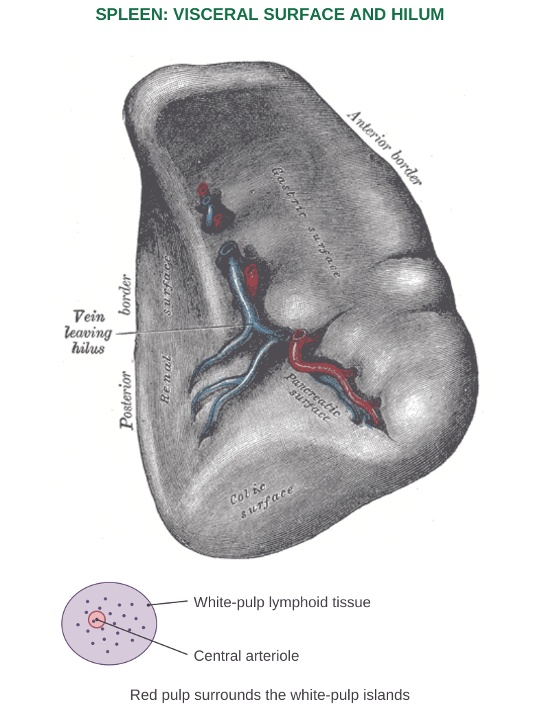
Diagram labels: Spleen: visceral surface and hilum. White-pulp lymphoid tissue; central arteriole. Red pulp surrounds the white-pulp islands.
Thymus
The thymus is lobulated, with a cortex and medulla in each lobule. Developing T cells undergo selection so that functional cells can recognise appropriate antigen presentation while strongly self-reactive populations are controlled.
Hassall corpuscles occur in the medulla. Thymic lymphoid tissue generally decreases after puberty, but this is not equivalent to loss of all T-cell immunity.
Tonsils and MALT
Tonsils survey material entering the pharyngeal region. Mucosa-associated lymphoid tissue protects mucosal surfaces; Peyer patches and other gut-associated tissues monitor intestinal antigens.
These lymphoid sites combine cellular organisation with their particular anatomical exposure.
3.18 Lymph Circulation and Drainage Territories
Lymph flows through initial capillaries, collecting vessels, nodes, larger trunks and ducts before returning to venous blood near the neck. Valves favour forward movement.
| Duct | Main drainage territory | Typical venous entry |
|---|---|---|
| Right lymphatic duct | Right head/neck, right upper limb and right thorax | Right venous angle |
| Thoracic duct | Remaining major territories, including both lower limbs and left upper body | Left venous angle |
The venous angle is the junction of the internal jugular and subclavian veins. Anatomy varies; the right-sided trunks may enter veins separately.
A cisterna chyli, when present, receives abdominal/lower-body lymph and continues as the thoracic duct.
Driving Forces
There is no single lymphatic pump equivalent to the heart. Skeletal-muscle movement, breathing-related pressure changes, nearby arterial pulsation and intrinsic collecting-vessel contractions promote flow.
One-way valves reduce backflow between collecting-vessel segments.
Intestinal Fat Transport
Lacteals within intestinal villi receive absorbed dietary lipid packaged into chylomicrons. Intestinal lymph travels through larger lymphatics to the thoracic duct and venous circulation.
Water-soluble nutrients often follow different absorption routes; not all intestinal nutrients enter lacteals.
3.19 Functions and Failures of Lymphatic Drainage
- Fluid balance: Return of interstitial water and escaped plasma proteins.
- Lipid absorption: Transport of chylomicrons from intestinal lacteals.
- Immune surveillance: Movement of antigens and immune cells to lymphoid sites.
- Host defence: Organised activation and expansion of immune populations.
Lymphoedema
Congenital abnormalities, obstruction, surgery, radiation injury or some infections may impair drainage. Protein-rich fluid accumulates, with persistent cases promoting tissue inflammation and fibrosis.
Distinguish a failure of lymph return from oedema caused primarily by reduced plasma protein.
Integrating Blood and Lymph
Blood delivers oxygen and substrates; microvascular exchange supplies tissue spaces; lymph recovers excess fluid and macromolecules.
Immune cells and mediators can use both networks, so inflammation changes not only cell behaviour but also fluid movement and drainage demand.
3.20 Inflammation: Introduction and Classification
Inflammation is a protective response of vascularised tissue to injury or harmful stimuli. It recruits plasma proteins and leukocytes to remove the cause, clear damaged material and prepare for repair.
It may occur without infection, for example after trauma, ischaemia or crystal deposition.
Cardinal Signs
| Sign | Main physiological basis |
|---|---|
| Redness, rubor | Increased local blood flow |
| Heat, calor | Increased flow and metabolic activity |
| Swelling, tumour | Fluid and cellular accumulation |
| Pain, dolor | Mediators and pressure on sensory structures |
| Loss of function | Pain, swelling or structural injury |
Acute Versus Chronic
| Feature | Acute | Chronic |
|---|---|---|
| Typical course | Rapid onset, shorter duration | Persistent or recurrent |
| Predominant cellular pattern | Often neutrophils early | Often macrophages and lymphocytes |
| Major tissue events | Vascular leakage and cell recruitment | Persistent injury plus attempts at repair |
Patterns vary with the stimulus. “Acute” does not automatically mean mild, and “chronic” does not simply mean an acute response lasting a few extra hours.
Morphological Patterns
- Serous inflammation: Produces relatively watery fluid.
- Fibrinous inflammation: Features fibrin-rich exudate.
- Suppurative inflammation: Produces pus with neutrophils and necrotic debris; an abscess is a localised collection.
- Ulcer: A surface defect caused by loss of tissue.
- Granulomatous inflammation: A distinctive chronic pattern with organised macrophage aggregates.
3.21 Acute Inflammation: Vascular Events
Resident cells recognise microbial products or tissue injury and release mediators. Arteriolar dilation increases flow, explaining redness and warmth. Changes in small-vessel permeability allow protein-rich fluid to leave the vascular space.
Permeability and Stasis
Endothelial gaps, direct endothelial injury or other barrier changes permit leakage. Loss of fluid from vessels raises the relative cellular concentration and slows flow, encouraging leukocyte margination.
Many permeability and recruitment events occur in postcapillary venules.
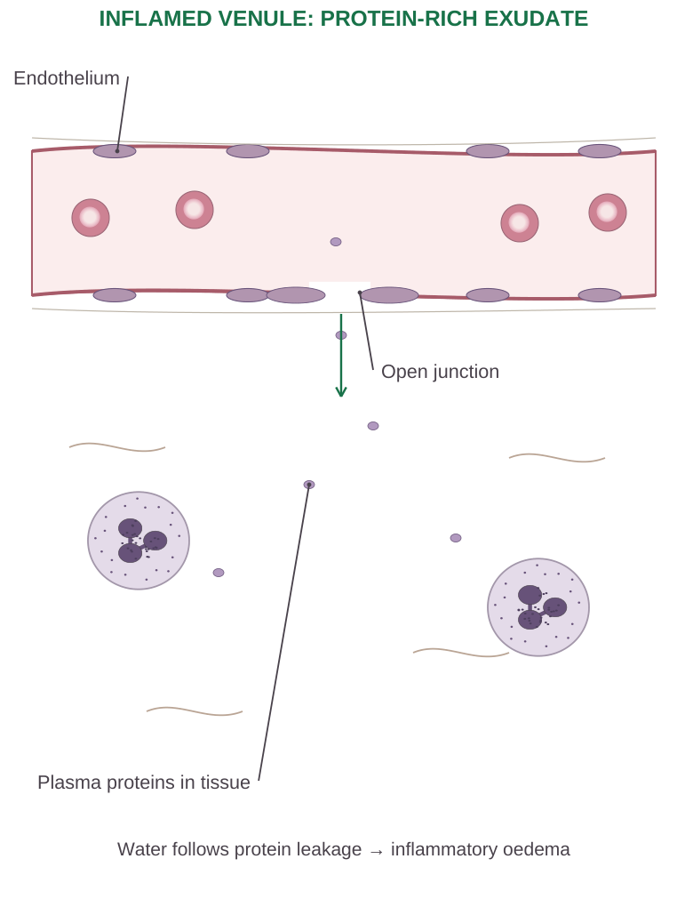
Diagram labels: Endothelium; open junction; plasma proteins in tissue. Water follows protein leakage → inflammatory oedema.
Exudate Versus Transudate
Exudate is generally protein-rich and associated with inflammatory permeability changes. Transudate is relatively protein-poor and results mainly from hydrostatic/oncotic imbalance without primary inflammatory barrier injury.
The appearance of fluid alone is insufficient to define its mechanism.
Benefit and Cost
Plasma proteins bring antibodies, complement and coagulation substrates. Swelling can dilute or contain harmful agents, but excessive oedema raises tissue pressure and may impair function.
A protective response can therefore become damaging if intense, misdirected or unresolved.
3.22 Leukocyte Recruitment and Phagocytosis
Leukocyte movement from blood to tissue requires successive changes in contact with the endothelium. Draw the process in relation to a vessel wall and an injury site, not as unrelated floating arrows.
Recruitment Sequence
- Step 1: Margination. Slower flow favours leukocytes near the vessel wall.
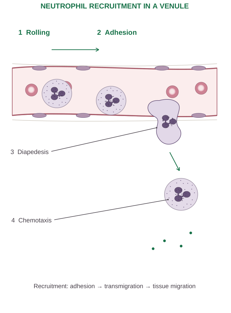
Diagram labels: Rolling; adhesion; diapedesis; chemotaxis. Recruitment: adhesion → transmigration → tissue migration.
- Step 2: Rolling. Selectin-mediated, transient attachments slow the cells.
- Step 3: Firm adhesion. Activated leukocyte integrins bind endothelial ligands such as ICAM/VCAM.
- Step 4: Transmigration. Cells cross the endothelial barrier, involving junctional molecules such as PECAM-1.
- Step 5: Chemotaxis. Cells move through tissue along gradients towards the injury.
Neutrophils often dominate early; monocytes/macrophages become prominent later. Timing varies by cause and tissue. Recruitment is directed cellular movement, not simply blood being poured out of the vessel.
Phagocytosis and Killing
Recognition and attachment are improved by opsonins such as IgG and complement fragments. Membrane pseudopods surround a target, forming a phagosome. Fusion with lysosomes produces a phagolysosome. Killing uses reactive oxygen species, enzymes and other mechanisms.
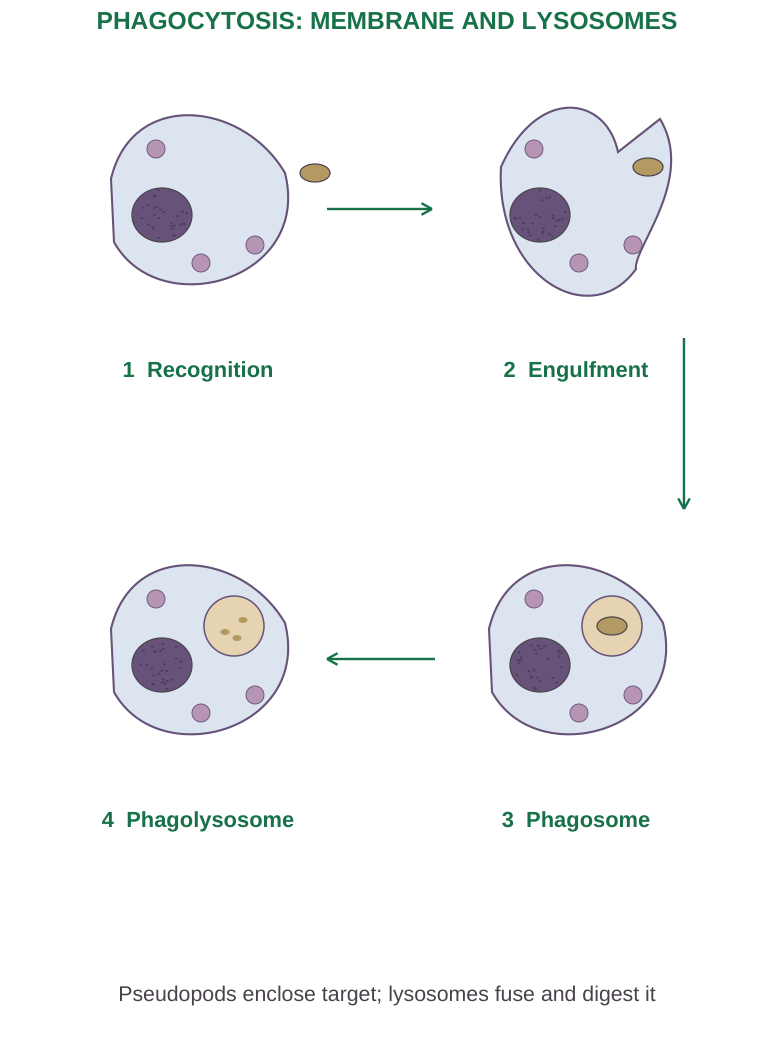
Diagram labels: Recognition; engulfment; phagosome; phagolysosome. Pseudopods enclose target; lysosomes fuse and digest it.
The NADPH oxidase respiratory burst generates reactive oxygen species; neutrophil myeloperoxidase supports formation of potent oxidants. Released enzymes and oxidants may also injure surrounding host tissue.
Macrophages clear debris and dying cells and help switch the response towards resolution.
3.23 Major Mediators of Inflammation
Mediators arise from cells or plasma-protein cascades. Some are preformed; others are synthesised after activation. Their effects depend on concentration, receptor expression and local context.
| Mediator | Principal teaching effects |
|---|---|
| Histamine | Rapid vasodilation/permeability changes |
| Prostaglandins | Vascular effects; PGE2 contributes to pain sensitisation and fever |
| Leukotrienes | LTB4 recruits/activates leukocytes; cysteinyl leukotrienes increase permeability and bronchoconstriction |
| Bradykinin | Pain, permeability and vasodilation |
| TNF and IL-1 | Endothelial activation; systemic inflammatory effects |
| Chemokines | Directed leukocyte recruitment |
| Complement | C3b opsonisation; C5a chemotaxis; C3a/C5a inflammatory effects |
| Nitric oxide | Vascular relaxation; microbial killing in relevant cells |
| Reactive oxygen species | Microbial killing and possible host injury |
Arachidonic-Acid Link
Membrane phospholipids supply arachidonic acid after phospholipase activation. Cyclooxygenase pathways form prostaglandins and thromboxanes; lipoxygenase pathways form leukotrienes and other products.
Thromboxane A2 also links platelet activation to haemostasis.
Systemic Responses
IL-1, TNF and other signals contribute to fever and acute-phase responses. The liver produces proteins such as CRP and increases fibrinogen. Leukocytosis may occur.
Severe systemic dysregulation can impair perfusion and organ function. CRP or ESR indicates inflammation but does not by itself identify a specific diagnosis.
3.24 Outcomes, Chronic Inflammation and Repair
An acute response may resolve completely, form an abscess, organise into fibrous tissue or persist as chronic inflammation. The outcome depends on the cause, tissue regenerative capacity, extracellular-matrix integrity and completeness of removal of the insult.
Resolution
The injurious cause is eliminated, recruitment declines, mediators are inactivated and dead cells are cleared. Fluid drains and surviving cells restore normal function.
Removal of dying neutrophils by macrophages helps terminate the response rather than automatically intensifying it.
Chronic Inflammation
Persistent infection, immune-mediated disease or prolonged exposure to harmful material can maintain inflammation. Macrophages, lymphocytes and plasma cells are often prominent.
Tissue destruction and repair occur together; cytokine signalling can maintain vascular changes, fibroblast activity and fibrosis.
Granulomatous Pattern
A granuloma contains an organised collection of activated macrophages, sometimes with multinucleated giant cells and surrounding lymphocytes. Some causes produce central necrosis; not every granuloma is caseating. A granuloma is not the same as granulation tissue.
Regeneration Versus Scar
Regeneration replaces lost tissue with the same functional cell type when cells and supporting matrix permit it. Scar formation replaces damage mainly with connective tissue when regeneration is insufficient.
Angiogenesis, fibroblast migration, collagen deposition and remodelling are central to repair.
Wound-Repair Connection
Haemostasis supplies an initial scaffold. Inflammation clears microbes and debris. Proliferation produces new epithelium, vessels and matrix. Remodelling reorganises collagen and strengthens the wound.
These phases overlap; repaired tissue may regain substantial strength without recovering all original architecture.
High-yield distinction: Granuloma: organised macrophage-rich inflammatory structure. Granulation tissue: vascular fibroblast-rich repair tissue. Similar words, different processes.
3.25 Anaemia: Mechanisms and Classification
Anaemia is reduced haemoglobin concentration relative to the appropriate reference threshold. Interpret age, sex, pregnancy, altitude and the laboratory’s context.
It reduces blood oxygen-carrying capacity; it is a finding requiring a cause, not a single disease.
Mechanistic Groups
| Mechanism | Examples |
|---|---|
| Reduced production | Iron/B12/folate deficiency, renal EPO deficiency, marrow failure |
| Increased destruction | Hereditary or acquired haemolysis |
| Blood loss | Acute haemorrhage or chronic bleeding |
Morphological Groups
Microcytic cells have a reduced mean corpuscular volume; iron deficiency and thalassaemia are important causes.
Macrocytic cells have increased MCV; B12/folate-related megaloblastic processes are key examples.
Normocytic anaemia occurs in several disorders, including acute blood loss and some chronic disease states.
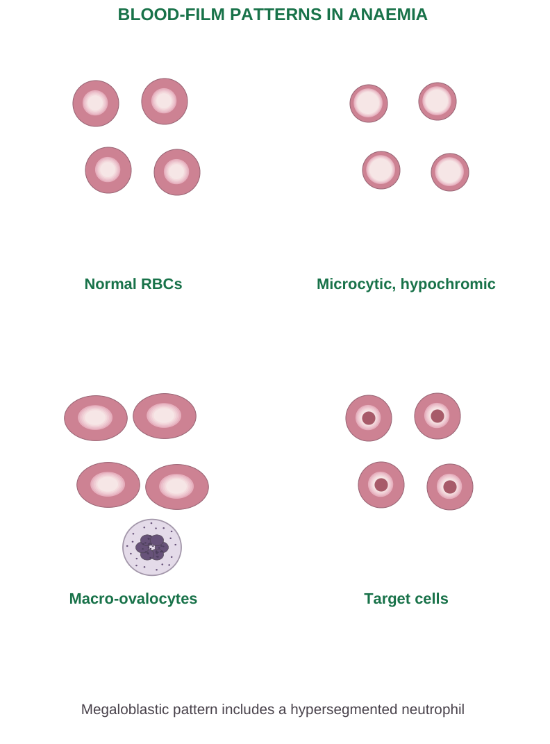
Diagram labels: Normal RBCs; microcytic, hypochromic cells; macro-ovalocytes; target cells. Megaloblastic pattern includes a hypersegmented neutrophil.
Basic Laboratory Reasoning
The CBC supplies Hb, haematocrit, RBC count and indices.
MCV = haematocrit (%) × 10 ÷ RBC count (million/µL)
This gives fL.
MCH = Hb (g/dL) × 10 ÷ RBC count
This gives pg.
RDW describes size variation; reticulocytes indicate marrow response. Smear appearance and biochemical tests refine the mechanism.
Shared Manifestations
Pallor, tiredness, exertional breathlessness, tachycardia and reduced exercise tolerance can result from inadequate oxygen delivery. Severity depends on concentration, speed of onset and comorbidity.
Jaundice favours increased RBC breakdown; neurological abnormalities suggest particular causes rather than all anaemias.
3.26 Iron-Deficiency Anaemia
Iron deficiency develops when iron supply or absorption is inadequate for requirements, or when losses exceed replacement. Causes include chronic blood loss, increased demand, poor intake and malabsorption.
Pathophysiology
- Step 1: Iron stores decline.
- Step 2: Iron available to marrow becomes inadequate.
- Step 3: Haemoglobin synthesis falls, limiting normal erythrocyte haemoglobinisation.
- Step 4: Established disease produces microcytic, hypochromic RBCs and reduced oxygen carriage.
Features and Investigations
The smear may show small pale cells, size variation and elongated pencil cells. Fatigue and pallor are common; some patients develop pica, glossitis or spoon-shaped nails.
Ferritin is typically low, transferrin saturation is reduced and iron-binding capacity often rises. Ferritin is also an acute-phase reactant, so inflammation can obscure iron deficiency with a normal or raised ferritin result.
Management Principle
Correct the underlying cause and replenish deficient iron using an appropriate clinical plan. Finding the source of blood loss matters as much as replacing iron.
Do not assume every microcytic anaemia needs iron; thalassaemia can be microcytic without deficiency.
3.27 Megaloblastic Anaemia: Vitamin B12 and Folate
Megaloblastic anaemia results from impaired DNA synthesis, commonly due to B12 or folate deficiency. Nuclear maturation lags behind cytoplasmic growth, producing large abnormal precursors and ineffective haemopoiesis.
Cells may die in marrow before reaching circulation.
Blood and Marrow Changes
The blood film often shows macro-ovalocytes and hypersegmented neutrophils. Severe disease can also lower leukocytes and platelets. Raised LDH and indirect bilirubin may reflect destruction of abnormal precursors.
Not every macrocytic anaemia is megaloblastic; other causes of macrocytosis exist.
| Feature | Vitamin B12 deficiency | Folate deficiency |
|---|---|---|
| Typical causes | Malabsorption, intrinsic-factor failure, inadequate intake | Poor intake, increased demand, malabsorption, some drugs |
| Neurological abnormalities | Can occur | Not the characteristic pattern |
| Homocysteine | May increase | May increase |
| Methylmalonic acid | Typically increases | Usually not increased from folate deficiency alone |
B12 Absorption and Pernicious Anaemia
Gastric intrinsic factor supports B12 absorption in the terminal ileum.
Pernicious anaemia is associated with autoimmune loss of intrinsic-factor-dependent absorption, often linked to gastric parietal-cell injury. It is one cause of B12 deficiency, not a synonym for all B12 deficiency.
Neurological Effects
B12 deficiency can affect peripheral nerves and spinal pathways, producing sensory changes, gait disturbance and other neurological findings. These can occur even when anaemia is not striking.
Folate may improve the blood picture while B12-related neurological injury persists; recognising B12 deficiency is therefore essential before treating an assumed isolated folate problem.
Core Principle
Identify the deficient vitamin and its cause, then replace appropriately. Management details depend on absorption, severity and clinical assessment; fixed self-treatment regimens are not required to explain the pathophysiology.
3.28 Sickle-Cell Anaemia and Sickle-Cell Disease
Sickle-cell disease arises from inherited haemoglobin variants involving HbS. Classical sickle-cell anaemia is HbSS; other genotypes, including HbSC and HbS/β-thalassaemia, can also produce sickle-cell disease.
Molecular Defect
HbS contains a β-globin substitution of valine for glutamate at position 6 in the conventional mature-chain numbering.
Deoxygenated HbS can polymerise, distorting RBCs and reducing deformability. Early sickling can reverse on oxygenation; repeated episodes damage membranes and promote persistent cell abnormality.
Two Major Consequences
Haemolysis: Damaged RBCs survive less well, causing anaemia, jaundice and compensatory marrow activity.
Vaso-occlusion: Rigid cells and adhesive interactions among RBCs, leukocytes, platelets and endothelium impair microvascular flow, producing ischaemia and pain.
The mechanism is more complex than a single curved RBC blocking a large artery.
Disease Manifestations
Painful episodes, acute chest syndrome, stroke, splenic dysfunction and other organ complications can occur. Repeated splenic injury may cause functional asplenia, increasing susceptibility to some infections.
Infection, dehydration and reduced oxygen availability can worsen disease, but not every episode has an obvious trigger.
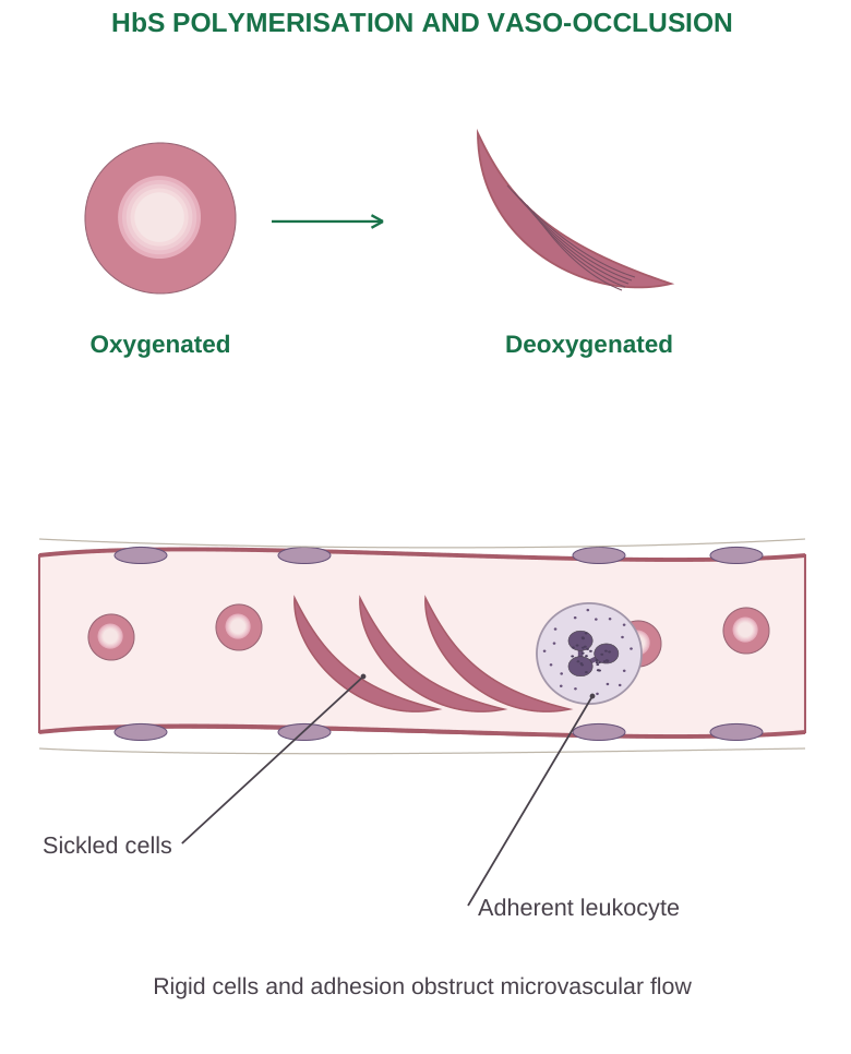
Diagram labels: Oxygenated; deoxygenated; sickled cells; adherent leukocyte. Rigid cells and adhesion obstruct microvascular flow.
Trait Versus Disease
Sickle-cell trait commonly means HbAS. It usually differs markedly from HbSS disease and is often asymptomatic under ordinary conditions, though special risks can occur in particular circumstances.
Do not describe every carrier as having severe sickle-cell anaemia.
Diagnosis and Management Concepts
Haemoglobin analysis and, when needed, genetic testing define the variant. Management combines prevention, complication-specific care and therapies that modify disease mechanisms.
Increasing HbF can reduce HbS polymerisation. The relevant learning point is the chain from molecular change to haemolysis and obstructed perfusion.
3.29 Thalassaemia: Reduced Globin-Chain Synthesis
Thalassaemias are inherited disorders with reduced production of α- or β-globin chains. The imbalance between paired chain types produces abnormal erythroid development, ineffective erythropoiesis and variable haemolysis.
Unlike HbS, the defining defect is chiefly quantity of chain synthesis, not the same structural substitution.
Alpha Thalassaemia
There are normally four α-globin gene copies. Loss or reduced function of increasing numbers generally increases severity: silent carrier, α-thalassaemia trait, HbH disease and severe α-chain deficiency with Hb Bart’s hydrops fetalis.
Genotype and whether affected genes are on the same chromosome matter for inheritance risk.
Beta Thalassaemia
β0 denotes absent β-chain production from an affected allele; β+ denotes reduced production.
Clinical patterns range from trait to severe transfusion-dependent disease. Historical “major/intermedia/minor” descriptions are useful, but modern classification also considers transfusion requirements.
Mechanism and Consequences
Unpaired chains injure developing erythroid cells. Marrow activity increases, but many precursors fail to mature effectively.
Severe disease can produce marrow expansion, splenomegaly and skeletal effects. Iron loading may result from repeated transfusion and, in some forms, increased intestinal absorption.
Laboratory Distinction
Microcytosis can be disproportionate to the Hb reduction. Target cells and other abnormalities may appear; an RBC count can remain relatively preserved in trait.
Haemoglobin analysis helps identify β-thalassaemia patterns, whereas some α-thalassaemia traits require different evaluation. Iron deficiency may coexist and complicate interpretation.
| Feature | Iron deficiency | Thalassaemia trait |
|---|---|---|
| Main defect | Inadequate iron for haem synthesis | Reduced globin-chain synthesis |
| Ferritin | Often low | Usually not low unless iron deficient |
| RBC count pattern | Often reduced as disease advances | May be relatively preserved |
| Specific evaluation | Iron status and cause of deficiency | Haemoglobin/genetic assessment as appropriate |
Do not prescribe iron merely because cells are small. Identify deficiency before assuming replacement will correct the disorder.
3.30 Hereditary and Acquired Anaemia
“Hereditary” and “acquired” describe origin; they do not replace morphological or mechanistic classification. A hereditary disorder can be haemolytic or production-related, and acquired disorders also have several mechanisms.
| Origin | Examples | Main mechanism |
|---|---|---|
| Hereditary membrane defect | Hereditary spherocytosis | Less deformable RBCs removed by spleen |
| Hereditary enzyme defect | G6PD deficiency | Reduced defence against oxidative injury |
| Hereditary haemoglobin defect | HbS and thalassaemia | Structural variant or reduced chain synthesis |
| Acquired immune haemolysis | Autoimmune haemolytic anaemia | Antibody-associated RBC destruction |
| Acquired mechanical injury | Microangiopathic fragmentation | RBC damage in abnormal microcirculation |
| Acquired marrow suppression | Aplastic anaemia and marrow injury | Reduced production, often multiple cell lines |
| Chronic inflammation/renal disease | Anaemia of inflammation; low EPO | Iron restriction/production impairment |
Haemolytic Pattern
Premature RBC destruction may raise reticulocytes, LDH and unconjugated bilirubin and lower haptoglobin, depending on the process.
Intravascular destruction occurs within blood; extravascular destruction is mainly macrophage-mediated. Reticulocytes may fail to rise if production is also impaired.
Selected Mechanisms
Hereditary spherocytosis changes membrane support and favours splenic clearance.
G6PD deficiency reduces generation of NADPH needed for antioxidant protection; oxidative stress can trigger episodes.
Autoimmune haemolysis involves antibodies, sometimes with complement.
Schistocytes suggest fragmentation, whereas spherocytes suggest reduced membrane surface relative to volume, but no isolated smear finding supplies the entire diagnosis.
Anaemia of Inflammation
Inflammatory signalling increases hepcidin, limiting iron export through ferroportin and making iron less available to erythropoiesis.
Reduced production and shortened RBC survival can contribute. Iron can be present in stores but poorly accessible; this differs from simple depletion of total stores.
3.31 Haemophilia
Haemophilia A is associated with factor VIII deficiency; haemophilia B with factor IX deficiency.
Classical inherited A and B are usually X-linked, though females can also have clinically important disease or bleeding. Factor XI deficiency has a different inheritance pattern and is sometimes called haemophilia C.
Pathophysiology
VIIIa and IXa cooperate in the intrinsic tenase complex to activate factor X efficiently. Deficiency reduces the thrombin burst and stable fibrin formation.
An initial platelet plug can form, but fibrin reinforcement is inadequate; bleeding may be prolonged or recur.
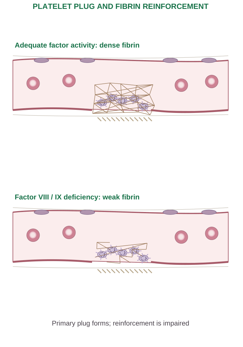
Diagram labels: Adequate factor activity: dense fibrin. Factor VIII/IX deficiency: weak fibrin. Primary plug forms; reinforcement is impaired.
Typical Pattern
Deep-muscle bleeding, haemarthroses and delayed bleeding after injury are important features. Repeated joint bleeding can damage synovium and cartilage.
Platelet number and primary platelet function are generally unaffected by isolated haemophilia.
| Investigation | Typical uncomplicated A/B pattern |
|---|---|
| Platelet count | Normal |
| PT | Usually normal |
| aPTT | Often prolonged; mild disease may escape screening |
| Factor assay | Reduced VIII or IX activity |
Inherited Versus Acquired
Acquired haemophilia commonly involves an inhibitor against factor VIII and differs from inherited X-linked disease. Inhibitors can also develop during treatment of inherited haemophilia.
Management targets the deficient factor or bypasses/modifies the coagulation defect under specialist care; the basic mechanism is defective fibrin reinforcement, not absence of all blood clotting.
3.32 Worked Cases and Rapid Revision
Case 1: Adequate Saturation, Reduced Carriage
A patient has low Hb but normal pulse-oximeter saturation. The saturation describes the occupied fraction of available sites, not the total haemoglobin amount. Anaemia can reduce oxygen content despite normal saturation.
Case 2: Distinguish Microcytic Mechanisms
Small pale RBCs plus low ferritin suggest iron depletion. Marked microcytosis with relatively preserved RBC count and non-depleted iron suggests a different possibility, including thalassaemia trait.
These patterns guide evaluation; they do not replace full testing.
Case 3: Identify an ABO Group
RBCs agglutinate with anti-A and anti-B reagents. The forward type suggests AB. Reverse typing and other required checks must be consistent before acceptance.
Lack of anti-D agglutination suggests D negativity under the validated testing method, not absence of every Rh antigen.
Case 4: Swelling After Node Removal
Regional swelling following interruption of lymphatic drainage reflects impaired return of tissue fluid and protein. The primary issue is drainage failure, not necessarily a fall in plasma albumin.
Case 5: Bleeding into a Joint
Recurrent haemarthrosis with normal platelet count, normal PT and prolonged aPTT suggests a coagulation-pathway defect such as haemophilia. Factor testing distinguishes VIII from IX deficiency and other causes.
Case 6: Interpret MCV
Haematocrit = 30%; RBC count = 3.0 million/µL.
MCV = 30 × 10 ÷ 3.0 = 100 fL
This is a size index; establish the laboratory interval and other findings before assigning a cause.
3.33 Examination Practice
Long-Answer Questions
- 1. Describe blood composition and explain the functions of its main constituents.
- 2. Explain haemopoiesis, erythroid maturation and EPO feedback with a labelled sketch.
- 3. Describe haemoglobin formation and connect haem/globin defects to anaemia.
- 4. Explain platelet-plug formation, coagulation and fibrinolysis.
- 5. Explain ABO and Rh grouping and component-specific transfusion compatibility.
- 6. Describe lymph formation, circulation, organs and functions with labelled diagrams.
- 7. Explain acute inflammatory vascular and cellular events and major mediators.
- 8. Compare iron-deficiency, megaloblastic, sickle-cell and thalassaemic mechanisms.
- 9. Explain hereditary/acquired anaemia and the pathophysiology of haemophilia.
Short-Answer Practice
- Plasma versus serum; haematocrit and buffy coat.
- Biconcave RBC structure and gas carriage.
- Neutrophil versus lymphocyte; platelets versus megakaryocytes.
- Primary versus secondary haemostasis.
- PT versus aPTT; fibrin versus fibrinogen.
- Afferent/efferent lymphatics; red/white splenic pulp.
- Exudate versus transudate; granuloma versus granulation tissue.
- B12 versus folate deficiency; sickle-cell trait versus disease.
Mechanism Comparison
| Condition | Defining disturbance |
|---|---|
| Iron deficiency | Inadequate haem synthesis substrate |
| B12/folate deficiency | Impaired DNA synthesis |
| Sickle-cell disease | HbS polymerisation with haemolysis/vaso-occlusion |
| Thalassaemia | Reduced globin-chain production |
| Haemophilia | Deficient fibrin reinforcement |
Quick Test: Answers Below
- 1. Which compartment contains fluid inside cells?
- 2. Which cells/fragments form the buffy coat?
- 3. Where do T cells mature?
- 4. What is the soluble precursor of fibrin?
- 5. Which blood group has A and B RBC antigens?
- 6. Which major lymphatic duct drains both lower limbs?
- 7. Which adhesion molecules support leukocyte rolling?
- 8. Which vitamin deficiency can cause characteristic neurological injury?
- 9. Which factor is deficient in haemophilia B?
Answers:
- 1. ICF.
- 2. Leukocytes and platelets.
- 3. Thymus.
- 4. Fibrinogen.
- 5. AB.
- 6. Thoracic duct.
- 7. Selectins.
- 8. B12.
- 9. IX.
Final Checklist
- Explain fluid compartments and solve a proportion calculation.
- Label normal RBCs, WBCs, platelets and erythroid maturation.
- Distinguish plasma, serum and packed RBCs.
- Trace tissue-factor initiation to cross-linked fibrin.
- Read ABO tables for the specified blood component.
- Trace lymph through a node to the venous angles.
- Locate cortex/paracortex/medulla and red/white pulp.
- Explain rolling, adhesion, diapedesis and chemotaxis.
- Compare all required anaemias by mechanism, morphology and distinguishing findings.
- Connect factor deficiency to deep-tissue bleeding.
Unit III Complete
Practise one labelled blood-cell plate, one lymph-node section and one mechanism-based disease comparison each revision session.
One-Minute Recall
- Blood: Plasma + RBCs + WBCs + platelets.
- Production: Marrow, growth factors and nutrients.
- Haemostasis: Adhesion → activation → aggregation → fibrin.
- Lymph: Tissue-fluid recovery, fat transport and immune surveillance.
- Inflammation: Vascular leakage + directed cell recruitment + resolution or repair.