Course Code: BP104T
Unit I Introduction
Introduction to the Human Body: Cellular Organization, Tissues and Cell Injury establishes the language and mechanisms needed to study anatomy, physiology and pathophysiology. It connects normal structure and function with adaptation, injury and cell death.
The unit moves from body organization and homeostasis to cells, transport, communication and tissues. It then explains why cells become injured, how injury develops and how cells respond.
Topics Covered – Complete Syllabus Map
| Required topic | Sections |
|---|---|
| Meaning and scope of the three disciplines | 1.1 |
| Levels of organization and body systems | 1.2-1.3 |
| Basic life processes | 1.3 |
| Anatomical terminology | 1.4-1.5 |
| Homeostasis and feedback components | 1.6 |
| Cell structure and functions | 1.7-1.9 |
| Transport across membranes | 1.10-1.11 |
| Cell division | 1.12-1.13 |
| Cell junctions | 1.14 |
| Cell communication and signalling forms | 1.15-1.16 |
| Tissue classification, structure, location and function | 1.17-1.22 |
| Causes and mechanisms of injury | 1.23-1.25 |
| Swelling, reversible and irreversible injury | 1.26 |
| Adaptation and dysplasia | 1.27-1.28 |
| Intracellular accumulation and cell death | 1.29-1.30 |
| Relevant medical terminology | 1.31 |
| Worked cases, revision and exam practice | 1.32-1.33 |
Learning Outcomes
- Relate anatomical structure to physiological function.
- Use standard terms for location, direction and body planes.
- Explain feedback and dynamic internal regulation.
- Identify major cellular structures and their functions.
- Compare membrane transport, cell division and junctions.
- Distinguish contact-dependent, paracrine, synaptic and endocrine signalling.
- Compare the four basic tissue groups with examples.
- Explain mechanisms and morphological outcomes of injury.
- Distinguish adaptation, dysplasia, accumulation and cell death.
Essential Vocabulary
| Term | Core meaning |
|---|---|
| Anatomy | Study of structure |
| Physiology | Study of normal function |
| Pathophysiology | Study of disturbed function in disease |
| Homeostasis | Dynamic regulation of the internal environment |
| Cell | Basic living structural and functional unit |
| Tissue | Organized cells and associated material serving functions |
| Adaptation | Change helping a cell cope with a demand |
| Injury | Cellular disturbance caused by a harmful influence |
| Necrosis | Injury-associated cell death with loss of membrane integrity |
| Apoptosis | Regulated cell dismantling with characteristic morphology |
Study Route
Learn the definition, inspect the diagram, compare similar concepts and explain the mechanism in your own words. Link every tissue to structure, location and function. Link every injury to cause, mechanism and outcome.
Central question: How does normal organization support function, and what changes when a demand exceeds the cell’s capacity to cope?
1.1 Anatomy, Physiology and Pathophysiology
Anatomy describes what the body is made of and how its parts are arranged. Physiology explains how those parts work. Pathophysiology explains how function becomes disturbed in disease or injury. Studying the three together makes mechanisms easier to understand.
Branches and Scope
| Area | Main focus | Example |
|---|---|---|
| Gross anatomy | Structures visible without microscopy | Heart chambers and major vessels |
| Microscopic anatomy | Cells and tissues | Epithelium and muscle fibres |
| Histology | Tissue organization | Lining of an intestine |
| Cytology | Cells | Nucleus and organelles |
| Regional anatomy | Structures within one region | Thorax |
| Systemic anatomy | Structures of a body system | Nervous system |
| Developmental anatomy | Changes during development | Formation of organs |
Structure-Function Relationship
A thin exchange surface supports diffusion; a thick protective layer resists abrasion; a muscle fibre contains machinery for contraction. Structure is therefore not a list of shapes but part of the explanation of function.
Pathophysiological Reasoning
Start with the normal function. Identify the disturbance and explain how it produces an altered function or manifestation. For example, inadequate oxygen delivery can reduce ATP production, impair ion pumps and contribute to cell swelling. This links a cause to a mechanism and an outcome.
Relevance to Pharmacy
Drug action depends on receptors, membranes, transport and tissue responses. Distribution depends partly on barriers and circulation. Injury can alter organ function and influence how the body handles medicines. These connections explain why foundational biology matters before studying therapeutics.
Worked comparison:
- Anatomy: a cell has a plasma membrane.
- Physiology: the membrane regulates exchange.
- Pathophysiology: severe membrane damage allows uncontrolled movement and leakage.
Examination rule: Do not define pathophysiology merely as “study of disease.” State that it concerns altered mechanisms and function; pathology also includes the causes and structural features of disease.
1.2 Levels of Structural Organization
The body has levels of organization, each building on the previous one. The levels are related, but the function of an organized whole is more than a collection of isolated parts.
Main Sequence
Chemical → cellular → tissue → organ → organ system → organism
Atoms combine into molecules; molecules contribute to organelles and cells; cells and extracellular material form tissues; multiple tissues form organs; organs cooperate in systems.
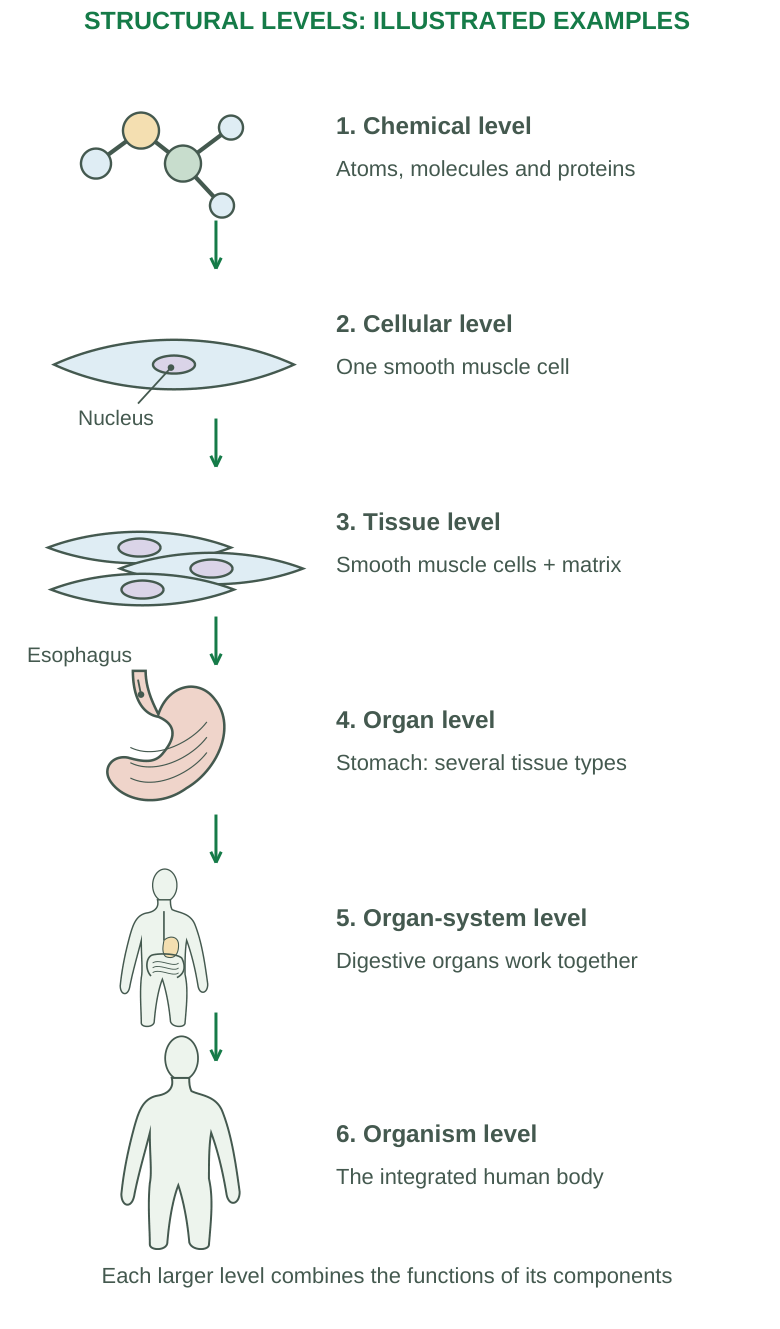
Diagram labels: Chemical level: atoms, molecules and proteins. Cellular level: one smooth muscle cell. Tissue level: smooth muscle cells + matrix. Organ level: stomach, several tissue types. Organ-system level: digestive organs work together. Organism level: the integrated human body. Nucleus; esophagus. Each larger level combines the functions of its components.
| Level | Example | Functional importance |
|---|---|---|
| Chemical | Water, ions, proteins and DNA | Reactions, structure and information |
| Cellular | Smooth muscle cell | A specialized living unit |
| Tissue | Smooth muscle tissue | Coordinated contraction |
| Organ | Stomach | Storage, mixing and digestion |
| System | Digestive system | Processing and absorption of nutrients |
| Organism | Human being | Integrated life processes |
Organs Contain Several Tissues
The stomach includes epithelium, connective tissue, muscle and nervous elements. Its function requires their cooperation. Calling an organ “a large tissue” misses this organization.
Extracellular Material
Cells interact with fluid and extracellular matrix. Matrix provides physical support and biochemical signals; extracellular fluid carries substances and contributes to the internal environment. Tissue includes these relationships, not only cell counts.
Clinical Connection
A molecular defect may disturb a cell, affect a tissue and impair an organ. Conversely, organ-level failure can change oxygen, nutrients or wastes in the cellular environment. The causal relationship can run through several levels.
Quick check: A group of similarly organized cells performing a related function is a tissue; an organized structure composed of different tissues is an organ. A single named tissue does not become an organ solely because it is large.
1.3 Body Systems and Basic Life Processes
Body systems cooperate to maintain life. The classification is useful for study; no system functions completely independently of the others.
| System | Major structures | Principal contribution |
|---|---|---|
| Integumentary | Skin and associated structures | Protection, sensation and temperature regulation |
| Skeletal | Bones, cartilage and joints | Support, protection, movement framework and mineral storage |
| Muscular | Skeletal muscles | Movement, posture and heat production |
| Nervous | Brain, spinal cord and nerves | Rapid coordination and information processing |
| Endocrine | Hormone-producing organs and cells | Chemical regulation of activity |
| Cardiovascular | Heart and blood vessels | Transport and circulation |
| Lymphatic / immune | Lymph vessels, nodes and lymphoid tissues | Fluid return and defence |
| Respiratory | Airways and lungs | Gas exchange |
| Digestive | Alimentary tract and accessory organs | Digestion and nutrient absorption |
| Urinary | Kidneys and urinary passages | Excretion and fluid/electrolyte regulation |
| Reproductive | Reproductive organs | Gamete production and reproduction |
Basic Life Processes
- Metabolism: All chemical reactions, including building molecules (anabolism) and breaking them down (catabolism).
- Responsiveness: Detecting and responding to change.
- Movement: Motion of the body, organs, cells or substances.
- Growth: Increase in size through cells or extracellular material.
- Differentiation: Development of specialized characteristics.
- Reproduction: Formation of new cells and, at organism level, a new individual.
Digestion, absorption, circulation and excretion support these processes.
Integration Example
During activity, skeletal muscle uses ATP, the circulation supplies oxygen and nutrients, respiration supports gas exchange and nervous/endocrine signals coordinate responses. Studying the organs separately must not hide this integration.
Memory point: A life process describes an activity; a body system describes organized structures contributing to activities. One process can involve several systems.
1.4 Anatomical Position and Directional Terms
Anatomical position is the standard reference: body upright, head and eyes forward, upper limbs at the sides, palms facing forward and feet directed forward. Right and left refer to the subject, not the observer.
Directional terms describe relationships between structures. Use them in anatomical position even when the person is lying or the limb has moved.
| Term | Meaning | Example |
|---|---|---|
| Superior / cranial | Toward the head | Chest is superior to abdomen |
| Inferior / caudal | Toward the lower end | Pelvis is inferior to abdomen |
| Anterior / ventral | Toward the front | Sternum is anterior to heart |
| Posterior / dorsal | Toward the back | Vertebral column is posterior to sternum |
| Medial | Toward the midline | Nose is medial to eyes |
| Lateral | Away from the midline | Ears are lateral to eyes |
| Proximal | Nearer the attachment or origin | Elbow is proximal to wrist |
| Distal | Farther from attachment or origin | Fingers are distal to elbow |
| Superficial | Nearer the surface | Skin is superficial to muscle |
| Deep | Farther from the surface | Bone is deep to skin |
| Ipsilateral | On the same side | Right arm and right leg |
| Contralateral | On opposite sides | Right arm and left leg |
Regional Language
- Head: cephalic.
- Neck: cervical.
- Chest: thoracic.
- Abdomen: abdominal.
- Pelvis: pelvic.
- Upper limb: arm or brachial region, forearm or antebrachial region, wrist or carpal region.
- Lower limb: thigh or femoral region, leg or crural region and ankle or tarsal region.
Position Terms
Supine: Lying face upward.
Prone: Lying face downward.
These describe body position rather than a new reference system for every directional term.
Exam trap: In anatomical terminology, arm usually means shoulder to elbow, and leg means knee to ankle. Everyday language often uses these words more broadly.
1.5 Body Planes, Cavities and Membranes
A plane is an imaginary flat section through the body. It helps describe relationships and interpret sections or imaging.
| Plane | Division produced |
|---|---|
| Sagittal | Right and left portions |
| Midsagittal / median | Equal right and left halves |
| Parasagittal | Unequal right and left portions |
| Frontal / coronal | Anterior and posterior portions |
| Transverse / horizontal | Superior and inferior portions |
| Oblique | Section at an angle to the main planes |
Major Cavities
Dorsal cavity: Cranial cavity containing the brain and vertebral canal containing the spinal cord.
Ventral cavity: Thoracic and abdominopelvic cavities. The diaphragm separates the thoracic cavity from the abdominal portion.
The thoracic cavity includes pleural cavities around the lungs and the mediastinum; the heart lies within the pericardial cavity in the mediastinum. The abdominopelvic cavity includes abdominal and pelvic portions without a complete physical partition between them.
Serous Membranes
A parietal layer lines a cavity wall; a visceral layer covers an organ surface. A small fluid-filled potential space helps reduce friction. Pleura relates to lungs, pericardium to the heart and peritoneum to abdominal/pelvic relationships.
Abdominal Regions
The four quadrants are right upper, left upper, right lower and left lower.
The nine-region scheme uses:
- Right/left hypochondriac and epigastric.
- Right/left lumbar and umbilical.
- Right/left iliac and hypogastric regions.
Use in Description
“The lesion is superficial and lateral to the midline” gives relationships. “It is on that side” does not. Identify the subject’s side and reference structure in an answer.
Quick check: A transverse cut does not divide front from back; that is a frontal plane. A sagittal cut need not pass through the exact midline.
1.6 Homeostasis and Feedback Systems
Homeostasis is dynamic regulation that keeps internal conditions within ranges compatible with function. It is not complete constancy or equilibrium. Variables fluctuate, and reference values can vary with time, activity and physiological circumstances.
Feedback Components
| Component | Role |
|---|---|
| Controlled variable | Condition being regulated, such as temperature |
| Stimulus / disturbance | Change affecting the variable |
| Receptor / sensor | Detects the relevant change |
| Afferent pathway | Carries information toward the control centre |
| Control centre | Integrates information and organizes a response |
| Efferent pathway | Carries output toward an effector |
| Effector | Produces the response |
| Feedback | Effect of the response on the variable or process |
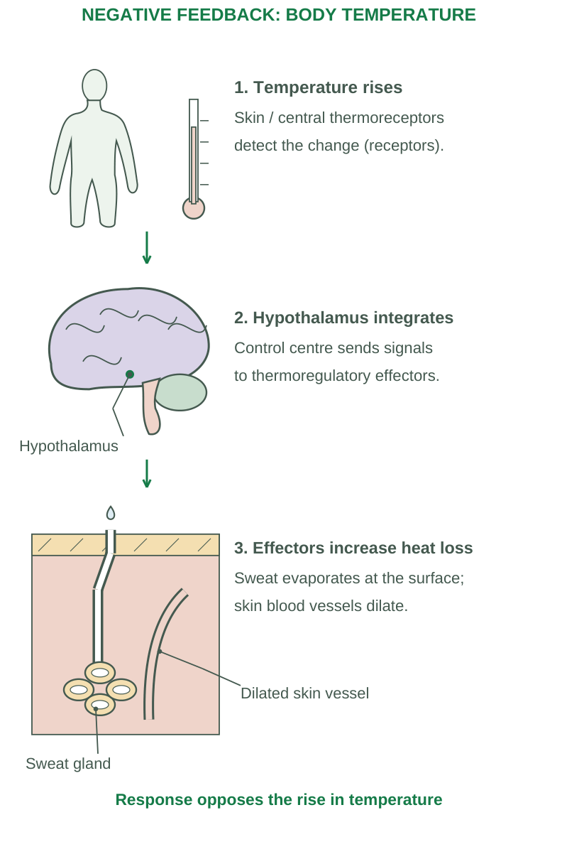
Diagram labels: Temperature rises: skin/central thermoreceptors detect the change. Hypothalamus integrates: control centre sends signals to thermoregulatory effectors. Effectors increase heat loss: sweat evaporates at the surface; skin blood vessels dilate. Hypothalamus; sweat gland; dilated skin vessel. Response opposes the rise in temperature.
Negative Feedback
The response opposes the original deviation. If temperature rises, relevant sensors and control mechanisms promote heat loss through sweating and skin blood flow. When the deviation is reduced, the response can diminish. “Negative” refers to the direction of feedback, not a harmful result.
Positive Feedback
The response reinforces the initial change and drives a process toward an endpoint. Cervical stretch during labour promotes signals that strengthen contractions, producing further stretch until delivery interrupts the cycle. Clotting also includes amplification, constrained by local and regulatory mechanisms.
Comparison
| Negative feedback | Positive feedback |
|---|---|
| Reduces a deviation | Amplifies a change |
| Supports regulation of many variables | Drives selected processes toward an endpoint |
| Example: temperature control | Example: labour contractions |
Failure and Disease
A disturbance may exceed the response capacity or damage the sensor, controller or effector. This can disrupt internal regulation. Feedback explains mechanisms; it does not mean every system has one simple controller or a fixed numerical set point.
Exam rule: Name the variable, sensor, control process, effector and feedback direction in an example. Listing “receptor-centre-effector” without explaining the response is incomplete.
1.7 The Cell: Structural and Functional Unit
A cell is the basic living unit of the human body. Human cells vary greatly in shape, size and specialization. A generalized nucleated cell helps organize study, but no single drawing represents every cell exactly.
Three Major Regions
- Plasma membrane: Boundary that regulates exchange and participates in communication.
- Cytoplasm: Cytosol and cellular structures outside the nucleus.
- Nucleus: Contains most cellular DNA and supports control of gene expression in nucleated cells.
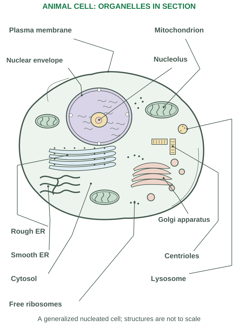
Diagram labels: Plasma membrane; mitochondrion; nuclear envelope; nucleolus; Golgi apparatus; rough ER; smooth ER; centrioles; cytosol; lysosome; free ribosomes. A generalized nucleated cell; structures are not to scale.
Function Depends on Specialization
Red blood cells are adapted for gas transport and lack a nucleus when mature. Muscle fibres contain contractile machinery. Secretory cells have substantial machinery for producing and exporting substances. Neurons have processes specialized for receiving and transmitting signals.
Cytosol and Organelles
Cytosol is the fluid component containing ions, soluble molecules and many enzymes. Organelles provide specialized compartments or structures. Compartmentalization separates reactions and supports efficient processing.
Cell Surface Specializations
Microvilli increase surface area for absorption. Motile cilia move fluid or material along a surface. A flagellum supports sperm movement. Microvilli and cilia differ in internal structure and function; they are not interchangeable hairs.
Cell Dependence on the Environment
Cells require suitable nutrients, oxygen for many metabolic activities, appropriate ions and removal of waste. The extracellular environment therefore connects cell biology to circulation, respiration, kidneys and other systems.
Exam point: Distinguish cytoplasm from cytosol and plasma membrane from a cell wall. Human cells have a plasma membrane but no rigid plant-type cell wall.
1.8 Organelles and Cytoskeleton
| Structure | Important features | Main functions |
|---|---|---|
| Nucleus | Double envelope with pores; chromatin | DNA storage, transcription and regulation |
| Nucleolus | Dense nuclear region without its own membrane | Ribosomal RNA production and subunit assembly |
| Ribosomes | RNA-protein particles, free or ER-bound | Protein synthesis |
| Rough ER | Membrane network with attached ribosomes | Processing proteins for secretion, membranes and some organelles |
| Smooth ER | Tubular membrane network without attached ribosomes | Lipid synthesis, detoxification and calcium handling in appropriate cells |
| Golgi apparatus | Stacked cisternae with receiving and shipping regions | Modification, sorting and packaging |
| Mitochondria | Double membrane; inner cristae | Oxidative ATP production and roles in cell death |
| Lysosomes | Acidic enzyme-containing compartments | Breakdown of material and recycling |
| Peroxisomes | Oxidative enzymes | Selected fatty-acid metabolism and peroxide handling |
| Centrosome | Microtubule-organizing region | Organization of microtubules and mitotic spindle |
Cytoskeleton
- Microfilaments, mainly actin, support shape, movement and contraction-related processes.
- Intermediate filaments provide mechanical stability and connect with certain junctions.
- Microtubules support intracellular transport, cilia and the mitotic spindle.
Protein Secretion Route
For many exported proteins:
Ribosome on rough ER → ER processing → transport vesicle → Golgi modification/sorting → secretory vesicle → plasma membrane release
Free ribosomes generally make proteins used in the cytosol or subsequently directed to certain other locations.
Important Distinctions
Ribosomes have no surrounding membrane. Lysosomes contain digestive enzymes; peroxisomes have different oxidative functions. Mitochondria produce much ATP under aerobic conditions, but glycolysis also produces ATP in the cytosol.
Memory aid: Rough ER builds and processes; Golgi modifies and sorts; vesicles deliver. These are linked functions rather than three separate names to memorize.
1.9 Plasma Membrane: Fluid Mosaic Model
The plasma membrane is a selectively permeable phospholipid bilayer containing proteins, cholesterol and associated carbohydrates. The fluid mosaic model describes a dynamic organization rather than a rigid sheet.
Components
| Component | Main contribution |
|---|---|
| Phospholipids | Hydrophilic heads face water; hydrophobic tails form the internal barrier |
| Cholesterol | Modulates fluidity, stability and permeability |
| Integral proteins | Span or enter the bilayer; channels, carriers, receptors and enzymes |
| Peripheral proteins | Associate with surfaces or other proteins |
| Carbohydrates | Cell recognition and surface interactions, mainly on the extracellular side |
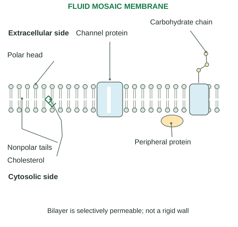
Diagram labels: Extracellular side; cytosolic side; polar head; nonpolar tails; channel protein; carbohydrate chain; cholesterol; peripheral protein. Bilayer is selectively permeable, not a rigid wall.
Selective Permeability
Small nonpolar molecules can cross the lipid region more readily than ions or large polar molecules. Water can move through lipid and, importantly in many cells, aquaporin channels. Specific transport proteins allow controlled movement of substances that do not easily cross the bilayer.
Membrane Functions
Boundary formation; transport; receptor-mediated communication; recognition; adhesion; linkage to cytoskeleton and matrix; and maintenance of ionic differences. These functions connect membrane structure with normal physiology and injury.
Gradients
A concentration gradient is a difference in substance concentration. An electrical gradient is a difference related to charge. For an ion, the electrochemical gradient combines both. Describing ion movement using concentration alone can be incomplete.
Why Membrane Damage Matters
Loss of selective permeability disturbs ions and water, permits leakage and can damage cellular processes. Severe membrane failure is central to irreversible injury, not merely a change in cell shape.
Quick check: A membrane protein is not automatically a transport protein. It may function as a receptor, enzyme, anchor or recognition molecule.
1.10 Passive Transport: Diffusion, Osmosis and Filtration
Passive transport does not directly require cellular metabolic energy for the transport step. Movement follows the relevant concentration, electrochemical, osmotic or pressure gradient.
Simple Diffusion
Substances move down a concentration gradient directly through the lipid bilayer. Small lipid-soluble molecules and gases can cross this region. Large polar substances and charged ions generally require suitable transport proteins instead of freely crossing the lipid barrier.
Facilitated Diffusion
Channels or carriers assist downhill movement. Channels provide selective routes for ions or water; many GLUT carriers transport glucose. Carrier-mediated transport is selective and saturable because carriers are limited. Protein involvement alone does not make transport active.
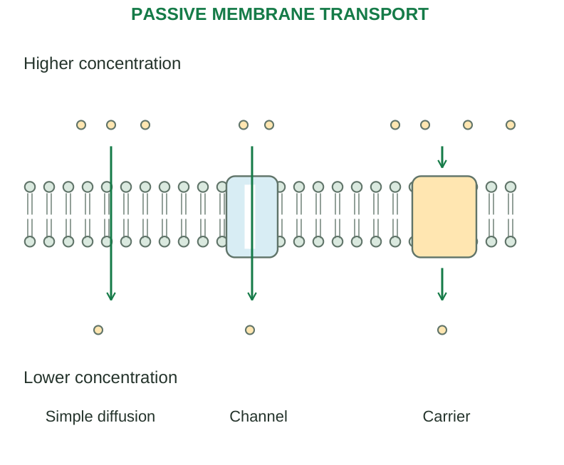
Diagram labels: Higher concentration; lower concentration; simple diffusion; channel; carrier. Passive membrane transport.
Osmosis
Water moves across a selectively permeable membrane toward the side with higher effective concentration of nonpenetrating solute.
- Isotonic: No sustained change in cell volume.
- Hypotonic: Water entry can swell the cell.
- Hypertonic: Water loss can shrink the cell.
Tonicity depends on solute permeability, not only total solute concentration.
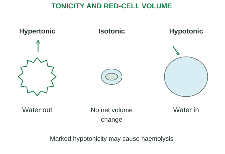
Diagram labels: Hypertonic: water out. Isotonic: no net volume change. Hypotonic: water in. Marked hypotonicity may cause haemolysis.
Filtration
A pressure difference drives water and small solutes through a barrier. Hydrostatic pressure and opposing forces influence movement. This is important in capillary exchange and renal filtration; it is not identical to diffusion.
Factors Affecting Diffusion
Gradient magnitude, surface area, permeability, molecule size and diffusion distance. A thin broad surface supports exchange; a thicker barrier reduces it when other factors remain comparable.
Conceptual relation: Rate of diffusion increases with area and gradient and decreases with barrier thickness. Use it to explain mechanisms, not as a universal numerical formula without units and assumptions.
Common error: Both facilitated diffusion and active transport can use carriers. Distinguish them by the direction of movement and energy coupling, not by presence of a protein alone.
1.11 Active Transport and Vesicular Movement
Active transport moves a substance against its electrochemical gradient by coupling transport to an energy source. Primary and secondary transport differ in the source used at the transport step.
Primary Active Transport
A pump directly uses energy, commonly from ATP hydrolysis. The sodium-potassium ATPase moves 3 sodium ions out and 2 potassium ions in per ATP in its standard cycle. It helps maintain gradients, cell volume and the electrical environment. It is electrogenic because charge movement is unequal.
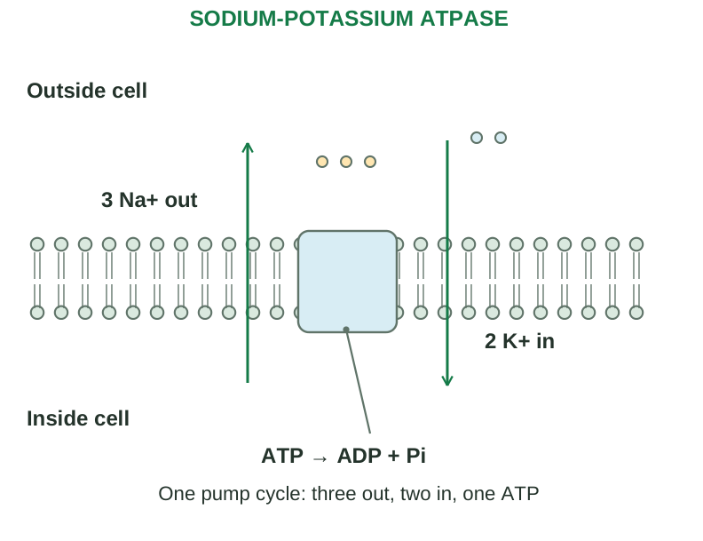
Diagram labels: Outside cell; inside cell; 3 Na+ out; 2 K+ in; ATP → ADP + Pi. One pump cycle: three out, two in, one ATP.
Secondary Active Transport
A downhill movement of one substance drives uphill movement of another. The coupled transporter does not directly hydrolyse ATP, but the driving gradient often depends on an ATP-consuming pump.
- Symport / cotransport: Coupled substances move in the same direction.
- Antiport / exchange: Coupled substances move in opposite directions.
Sodium-glucose cotransport is a classic example of secondary active transport; it differs from GLUT-mediated facilitated diffusion.
Vesicular Transport
- Endocytosis: Uptake through membrane-derived vesicles.
- Phagocytosis: Uptake of large particles by appropriate cells.
- Pinocytosis: Uptake involving extracellular fluid.
- Receptor-mediated endocytosis: Selective uptake following receptor binding.
- Exocytosis: Vesicle fusion releases contents and contributes membrane components.
- Transcytosis: Uptake on one side followed by transport and release on another side in suitable cells.
Comparison
| Mechanism | Energy / direction |
|---|---|
| Facilitated diffusion | Downhill through a carrier; no direct metabolic coupling |
| Primary active transport | Direct energy use for uphill movement |
| Secondary active transport | Energy of another gradient drives coupled movement |
| Vesicular transport | Energy-dependent membrane trafficking |
Injury link: ATP depletion impairs energy-dependent pumps and trafficking. Sodium and water accumulation can produce swelling; calcium disturbances can activate damaging enzymes.
1.12 Cell Cycle and Mitosis
The cell cycle is the ordered process of growth, DNA replication and division. Cycling cells pass through interphase and a division phase. Not all mature cells actively divide; some remain in a noncycling G0 state.
Phases
- G1: Growth and preparation.
- S: DNA replication.
- G2: Further preparation and checking before division.
- M: Mitosis and usually cytokinesis.
Interphase includes G1, S and G2 and is metabolically active, not a resting period.
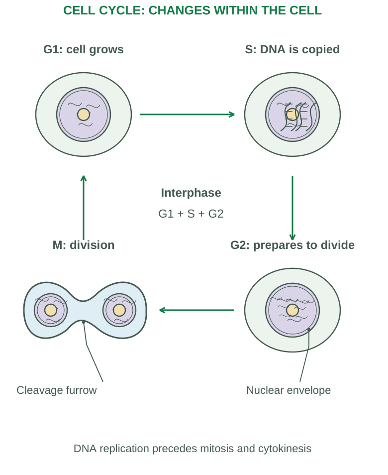
Diagram labels: G1: cell grows. S: DNA is copied. G2: prepares to divide. M: division. Cleavage furrow; nuclear envelope. Interphase: G1 + S + G2. DNA replication precedes mitosis and cytokinesis.
Mitosis Sequence
- Prophase: Chromosomes condense and the spindle develops.
- Prometaphase: Nuclear envelope breaks down and spindle microtubules attach to kinetochores.
- Metaphase: Chromosomes align near the equatorial plate.
- Anaphase: Sister chromatids separate.
- Telophase: Nuclei reform and chromosomes decondense.
Cytokinesis divides the cytoplasm, commonly through a contractile ring in animal cells. It overlaps with late mitosis rather than always beginning after all nuclear changes finish.
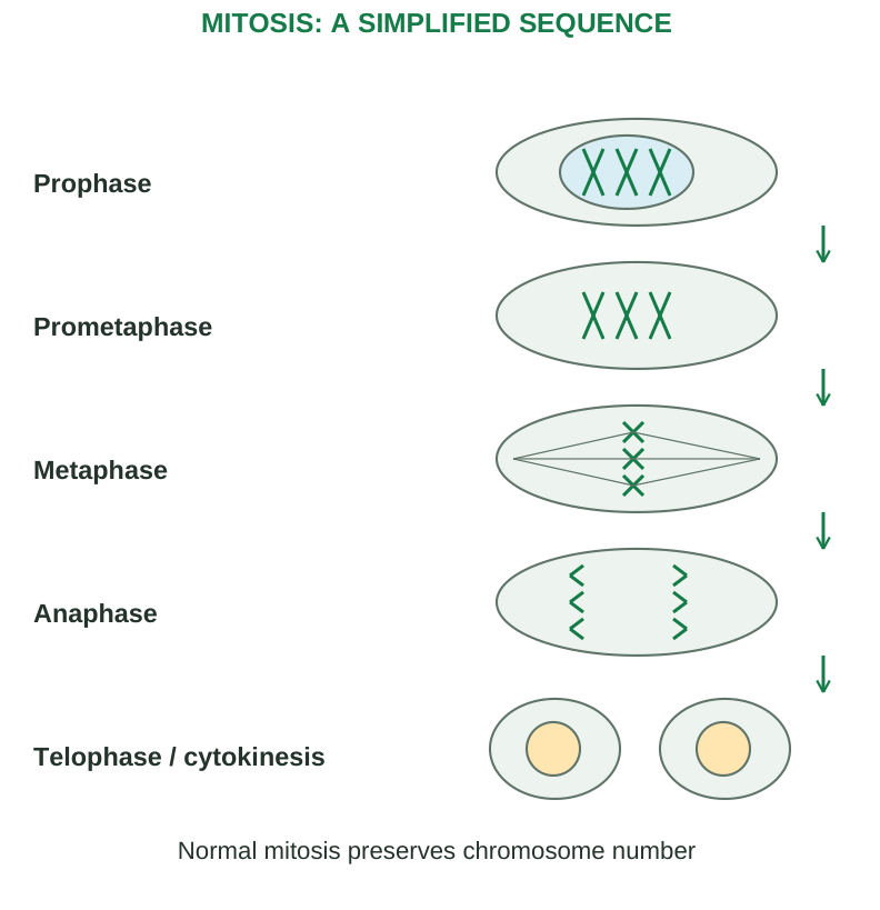
Diagram labels: Prophase; prometaphase; metaphase; anaphase; telophase/cytokinesis. Normal mitosis preserves chromosome number.
Result and Purpose
Mitosis generally produces two daughter cells with the same chromosome number as the original cell and closely matching genetic information. It supports growth, renewal and repair. Mutations can occur, so “always perfectly identical” is too absolute.
Cell-Cycle Control
Checkpoints and regulatory proteins help ensure conditions are suitable and DNA or spindle problems are addressed. Failure of regulation can contribute to abnormal proliferation. A checkpoint does not guarantee that every error is prevented.
Chromosome Language
DNA replication produces sister chromatids, but the chromosome number counted by centromeres is not simply doubled in S phase. In human somatic cells, the usual diploid number is 46; DNA content changes before division.
Memory aid: Prepare → copy → check → divide. For mitosis: condense, attach, align, separate and reform. State both nuclear division and cytokinesis when asked about complete cell division.
1.13 Meiosis and Comparison with Mitosis
Meiosis is a specialized division process that reduces chromosome number during formation of germ-line cells leading to gametes. It includes one round of DNA replication followed by two divisions.
Meiosis I
Homologous chromosomes pair during prophase I and may exchange corresponding segments through crossing over. Homologous pairs align at metaphase I; homologous chromosomes separate at anaphase I. Sister chromatids generally remain joined through this first division.
Meiosis II
No new S phase normally occurs between the two meiotic divisions. Sister chromatids separate during meiosis II. The resulting cells have a haploid chromosome complement. Human gametes normally have 23 chromosomes.
Comparison
| Feature | Mitosis | Meiosis |
|---|---|---|
| Main role | Growth, renewal and repair | Germ-line reduction division |
| Divisions | One | Two |
| Homologous pairing | Not a normal feature | Occurs in prophase I |
| Crossing over | Not a normal mitotic feature | Occurs between homologues in prophase I |
| Chromosome number | Usually maintained | Reduced to haploid |
| Genetic outcome | Closely matching daughters | Variation through recombination and assortment |
| Typical products | Two daughter cells | Four haploid products in the general scheme |
Variation
Crossing over and independent assortment contribute to genetic diversity. Fertilization combines haploid contributions and restores the diploid complement in the new individual.
Human Reproductive Qualification
The general four-product diagram does not mean four equally functional gametes in every process. Spermatogenesis and oogenesis differ; oogenesis involves unequal cytoplasmic divisions and polar bodies. Detailed reproductive physiology belongs to later study.
Exam trap: In meiosis I, homologues separate; in meiosis II, sister chromatids separate. DNA replication occurs before the first division, not before each division.
1.14 Cell Junctions: Barriers, Attachment and Communication
Cell junctions are specialized connections between cells or between a cell and extracellular material. Their functions include sealing, mechanical attachment and transfer of signals or small molecules.
| Junction | Main structure/function | Example of relevance |
|---|---|---|
| Tight junction | Protein-based seal limiting movement between adjacent cells | Epithelial barriers |
| Adherens junction | Cell-cell attachment linked mainly to actin | Tissue organization and force transmission |
| Desmosome | Strong cell-cell attachment linked to intermediate filaments | Skin and cardiac tissue mechanical strength |
| Hemidesmosome | Cell attachment to basement membrane through intermediate filaments | Basal epithelial anchoring |
| Gap junction | Channels allowing ions and small molecules between cells | Coordinated electrical activity |
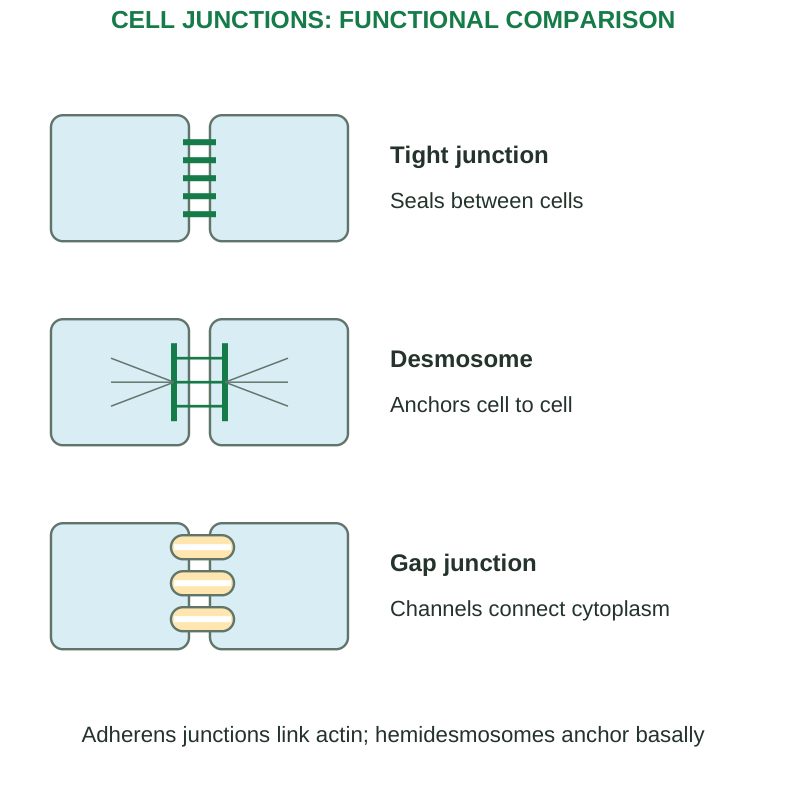
Diagram labels: Tight junction: seals between cells. Desmosome: anchors cell to cell. Gap junction: channels connect cytoplasm. Adherens junctions link actin; hemidesmosomes anchor basally.
Tight Junctions and Polarity
They restrict paracellular passage and help maintain differences between apical and basolateral membrane regions. They are selective barriers, not universal completely watertight walls.
Desmosomes Versus Hemidesmosomes
A desmosome connects cell to cell. A hemidesmosome connects cell to basement membrane. The shared name does not mean they are half-open communication channels.
Gap Junctions
Aligned channel complexes connect adjacent cytoplasms. They allow selected small substances, including ions, to pass; they do not normally transport whole organelles or large proteins. Their conductance and opening can be regulated.
Function in Injury
Damage to junctions can weaken a barrier or attachment, affecting permeability or tissue integrity. This differs from damage to an individual cell’s plasma membrane, although both can occur together.
Memory aid: Tight seals, anchoring holds, gap communicates. Then identify the specific anchoring partner and cytoskeletal connection for a complete answer.
1.15 General Principles of Cell Communication
Cell communication coordinates activity through signals and responses. A chemical signal influences a target only if the appropriate receptor and response machinery are present. Distribution of the signal alone does not make every cell respond identically.
General Sequence
Signal production → delivery → receptor binding → transduction → cellular response → regulation or termination
Different cells may interpret the same signal differently because receptors and downstream pathways differ.
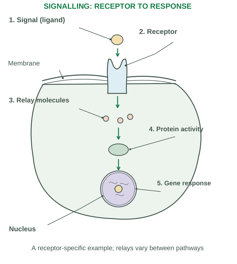
Diagram labels: Signal (ligand); receptor; membrane; relay molecules; protein activity; gene response; nucleus. A receptor-specific example; relays vary between pathways.
Receptor Location
Cell-surface receptors detect many signals that do not readily cross the plasma membrane. They can couple to ion channels, G proteins or enzyme-related pathways.
Intracellular receptors detect suitable signals that enter the cell, including many steroid hormone signals, and can influence gene expression.
Transduction
Binding is converted into intracellular events. These may include ion movement, activation of enzymes, second messengers or phosphorylation cascades. Common second messengers include cyclic AMP, calcium and IP3; not every pathway uses all of them.
Response and Amplification
Responses may alter secretion, metabolism, contraction, transport, gene expression, growth or survival. A pathway can amplify a small initiating signal. Amplification is regulated and does not imply limitless activity.
Ending the Signal
Signal removal, receptor desensitization, messenger breakdown and reversal of downstream changes help limit the response. Continued exposure can alter sensitivity. Without termination, a signal may produce inappropriate or prolonged effects.
Distinguish Communication Terms
A ligand is a molecule that binds a receptor. A receptor detects a signal; a second messenger relays it inside the cell. A hormone is defined by its signalling role, not by being necessarily a protein.
Examination rule: Explain how a target is selected and how receptor binding becomes a response. “Cells send messages” is a description, not a mechanism.
1.16 Forms of Intercellular Signalling
The syllabus requires four forms. Compare them by delivery route, distance and target relationship, not solely by whether the signal acts quickly.
| Form | How the signal reaches its target | Typical example |
|---|---|---|
| Contact-dependent / juxtacrine | A membrane-bound signal interacts with an adjacent cell’s receptor | Contact-mediated developmental or immune signalling |
| Paracrine | Released mediator acts locally on nearby cells | Local growth-factor effects |
| Synaptic | Neuron releases neurotransmitter across a specialized synaptic cleft | Communication with a neuron, muscle or gland |
| Endocrine | Hormone enters circulation and reaches distant targets | Insulin acting on appropriate tissues |
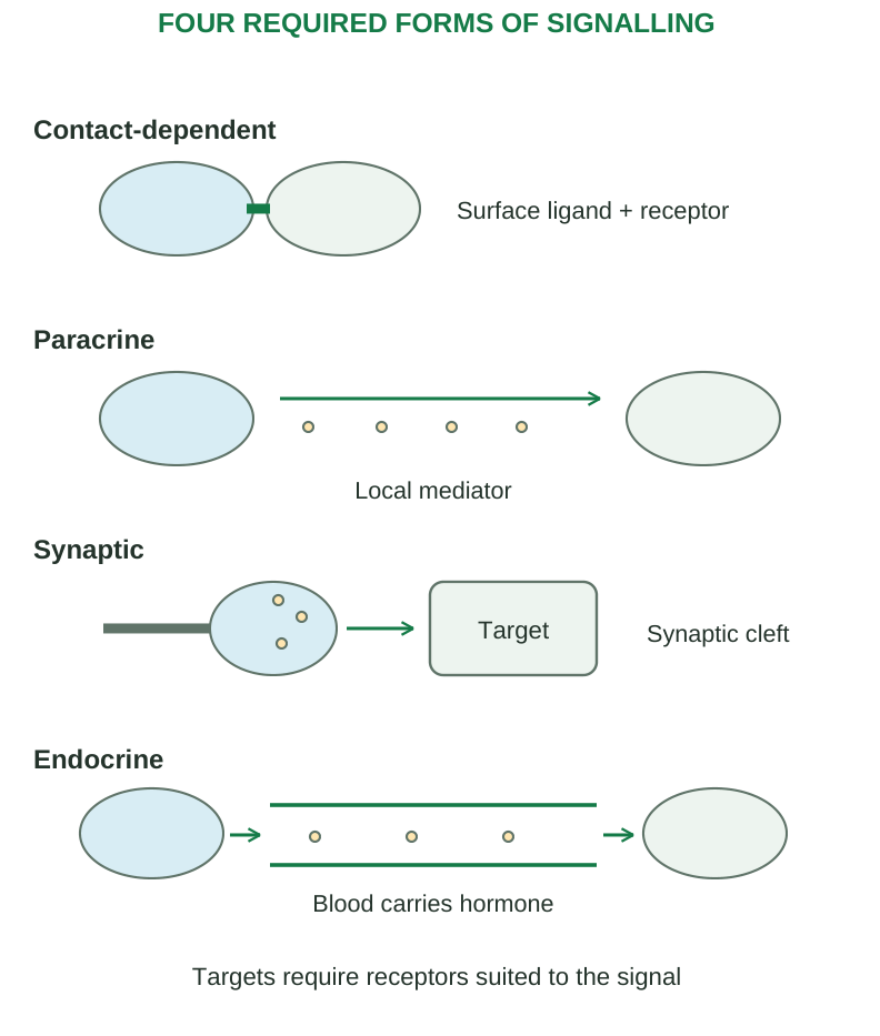
Diagram labels: Contact-dependent: surface ligand + receptor. Paracrine: local mediator. Synaptic: target and synaptic cleft. Endocrine: blood carries hormone. Targets require receptors suited to the signal.
Contact-Dependent
Cells need suitable close contact. The signalling ligand remains associated with the source membrane in the classic form. Do not confuse this with a gap junction, where a direct channel connects cytoplasms.
Paracrine
Signals diffuse locally and are limited by uptake, breakdown and other controls. “Local” does not mean that every nearby cell responds; receptor expression still determines responsiveness.
Synaptic
An electrical signal travels along the axon, and a chemical neurotransmitter is released at an appropriate terminal. The specialized target relationship supports precise communication. Synaptic signalling has a local chemical component but is conventionally discussed separately because of neuronal delivery and synaptic specialization.
Endocrine
The circulation carries the hormone beyond the immediate neighbourhood. Target cells must have relevant receptors. The speed and duration depend on the hormone and pathway; endocrine signalling is not always slow.
Additional Term: Autocrine
A released signal acts on the source cell or cells of the same relevant population. It is useful for comparison but does not replace any of the four required forms.
Quick comparison: Touch, nearby, synapse, bloodstream. Explain the route behind each word and include receptor specificity.
1.17 Four Basic Tissue Groups
Tissues are organized cells and associated extracellular material that perform related functions. The four broad groups are epithelial, connective, muscular and nervous tissues. Organs usually combine them.
| Tissue | Organization | Main contribution |
|---|---|---|
| Epithelial | Closely arranged cells, little intervening matrix | Covering, lining, secretion and absorption |
| Connective | Cells within varying extracellular matrix | Support, binding, transport and defence |
| Muscular | Cells specialized for contraction | Movement and force |
| Nervous | Neurons and supporting glial cells | Information processing and coordination |
Structure, Location and Function
A tissue answer should connect all three. “Simple squamous epithelium” identifies structure; “alveolar lining” identifies a location; “supports gas exchange” explains a function. A long definition with no location misses part of the syllabus.
Extracellular Matrix
Matrix contains ground substance and fibres in varying amounts. In connective tissue, its composition strongly influences function. Mineralized matrix supports bone rigidity; a fluid matrix enables blood transport.
Tissue Repair Potential
Cell renewal varies. Some populations divide frequently, others can re-enter the cycle when needed, and many highly specialized cells have limited replacement capacity. Matrix integrity, blood supply and severity of injury influence repair.
Membranes at Tissue Level
An epithelial membrane includes epithelium and underlying connective tissue. Mucous membranes line passages communicating with the exterior. Serous membranes cover suitable cavity surfaces. The cutaneous membrane is the skin. Synovial membranes are connective-tissue-based and are not a typical epithelium on a basement membrane.
Exam link: A barrier function suggests tightly organized epithelial cells; a support function suggests connective matrix; contraction suggests muscle; rapid signalling suggests nervous tissue. These are starting clues, not complete microscopic diagnoses.
1.18 Epithelium: General Features
Epithelium covers surfaces, lines passages and forms much glandular tissue. Its closely packed cells are arranged into sheets with specialized junctions and relatively little extracellular material between them.
Structural Features
- Polarity: Apical surface faces a lumen or exposed space; basal surface relates to basement membrane; lateral surfaces contact neighbours.
- Basement membrane: Provides support and anchoring and separates epithelium from underlying connective tissue.
- Avascularity: Epithelium generally lacks its own blood vessels; nutrients usually arrive by diffusion from underlying connective tissue or appropriate surface supply.
- Innervation and renewal: Many epithelia are innervated and renew actively, with variation between types.
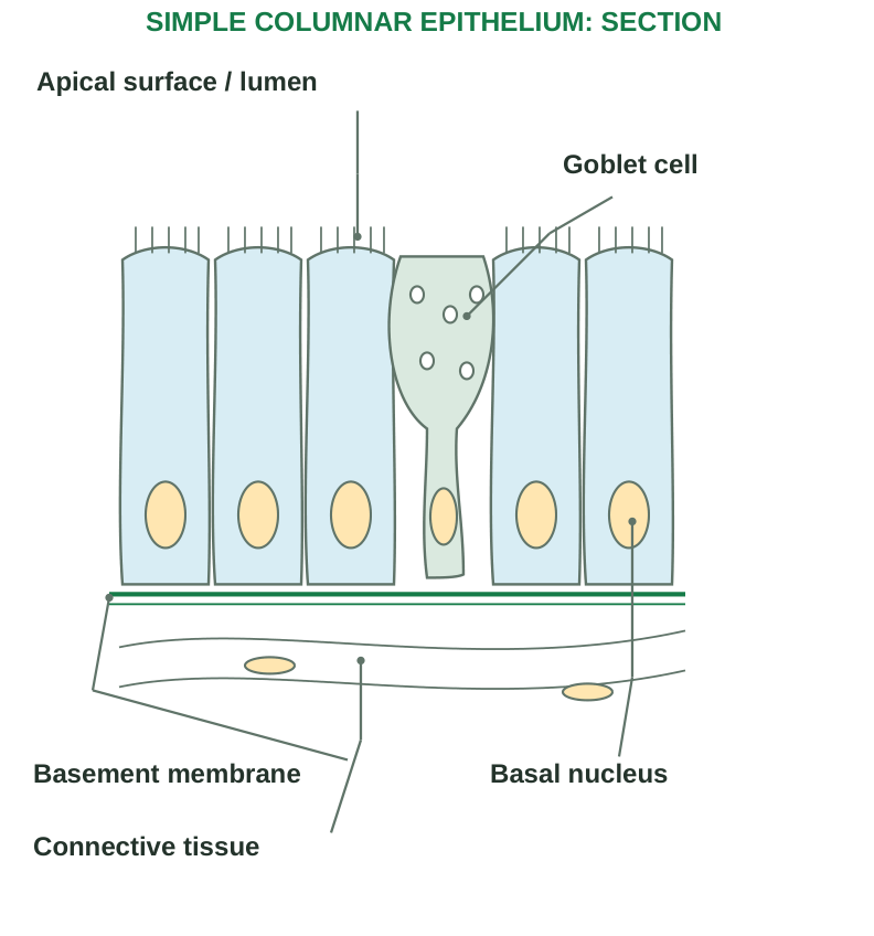
Diagram labels: Apical surface/lumen; goblet cell; basement membrane; basal nucleus; connective tissue.
Functions
Protection, absorption, secretion, filtration, exchange and sensation. A tissue’s structure should match its task: multiple layers resist abrasion; a thin layer supports exchange; surface specializations help absorption or movement of material.
Classification Rules
- Simple: One layer.
- Stratified: Multiple layers.
- Pseudostratified: All cells contact the basement membrane, but nuclei lie at different levels and not all cells reach the free surface.
- Squamous: Flattened.
- Cuboidal: Roughly as tall as wide.
- Columnar: Taller than wide.
Stratified epithelia are named mainly by the cells at the surface, not the deepest layer.
Specializations
Microvilli increase surface area. Motile cilia move material. Goblet cells secrete mucus in suitable epithelia. Keratinized surface layers contribute to a protective barrier.
Key distinctions: Pseudostratified does not mean truly multilayered. The basement membrane is extracellular support, not another living epithelial cell layer. Epithelium is not limited to skin.
1.19 Epithelial Types and Glands
| Type | Salient structure | Example location | Main function |
|---|---|---|---|
| Simple squamous | Thin single layer of flat cells | Alveoli, vessel endothelium, serosal mesothelium | Exchange, lining and reduced friction |
| Simple cuboidal | Single layer of cuboidal cells | Many kidney tubules and small gland ducts | Absorption and secretion |
| Simple columnar | Single layer of tall cells | Intestinal lining | Absorption and secretion |
| Ciliated columnar | Tall cells with apical cilia | Parts of uterine tubes | Movement of material |
| Pseudostratified ciliated columnar | Different nuclear levels, often goblet cells | Much of tracheal lining | Mucus production and transport |
| Stratified squamous, keratinized | Multiple layers with keratinized surface | Epidermis | Abrasion resistance and barrier |
| Stratified squamous, nonkeratinized | Multiple layers, moist living surface | Oesophagus and parts of oral cavity | Protection against abrasion |
| Stratified cuboidal / columnar | Uncommon multilayered duct lining | Selected larger ducts | Protection and secretion-related roles |
| Transitional / urothelium | Variable surface-cell shape with distension | Bladder and other urinary passages | Stretching and urine barrier |
Endocrine Versus Exocrine Glands
Endocrine: No duct for the secretion; products enter interstitial fluid and generally the circulation.
Exocrine: Release through a duct or directly onto an epithelial surface. Unicellular exocrine glands include suitable goblet cells; multicellular glands have organized secretory portions.
Secretion Mechanisms
- Merocrine: Exocytosis.
- Apocrine: Release involving an apical portion.
- Holocrine: Whole-cell breakdown contributes to secretion.
These terms describe secretion mechanisms, not simply the shape of a gland.
Study Method
For each type, recall layer number, surface shape or specialization, one representative location and the structure-function link. Avoid memorizing a location without knowing why the tissue fits it.
1.20 Connective Tissue: Matrix, Cells and Types
Connective tissue supports, binds, protects, stores, transports and participates in defence. Cells are distributed within a matrix whose quantity and composition vary widely.
Components
- Cells: Fibroblasts, adipocytes and various defence-related cells depending on the tissue.
- Fibres: Collagen provides tensile strength; elastic fibres support stretch and recoil; reticular fibres form delicate supporting networks.
- Ground substance: Hydrated material surrounding cells and fibres.
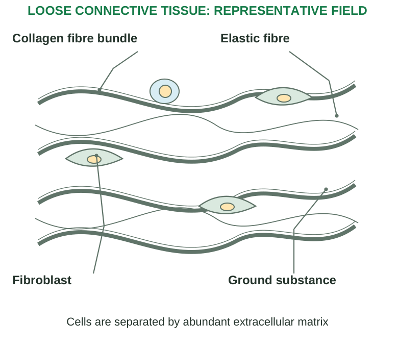
Diagram labels: Collagen fibre bundle; elastic fibre; fibroblast; ground substance. Cells are separated by abundant extracellular matrix.
| Type | Salient structure | Example location | Function |
|---|---|---|---|
| Loose areolar | Mixed fibres and cells with space for fluid | Beneath many epithelia | Packing, support and diffusion environment |
| Adipose | Lipid-storing adipocytes | Subcutaneous and selected organ-associated areas | Energy storage, cushioning and endocrine roles |
| Reticular | Fine supporting network | Supporting framework in suitable lymphoid organs | Supports cells |
| Dense regular | Parallel collagen bundles | Tendons and many ligaments | Strength along a dominant direction |
| Dense irregular | Interwoven collagen | Dermis and organ capsules | Resists forces from varied directions |
| Hyaline cartilage | Smooth-appearing matrix with chondrocytes in lacunae | Articular surfaces, tracheal supports | Support and low-friction surfaces |
| Elastic cartilage | Matrix rich in elastic fibres | External ear | Flexible support |
| Fibrocartilage | Strong collagen-rich matrix | Intervertebral discs | Resistance to compression and tension |
| Bone | Mineralized matrix with organized cells | Skeleton | Support, protection and mineral storage |
| Blood | Cells suspended in plasma | Vessels | Transport, defence and haemostasis |
Important Qualifications
Cartilage is avascular and its cells depend on diffusion; repair can be limited. Bone is vascular and remodels. Blood remains connective tissue despite its fluid matrix. Dense regular tissue is not strong equally in every direction.
Contrast: Epithelium emphasizes closely joined cells; connective tissue emphasizes the properties of its matrix. Both can be present in the same organ.
1.21 Muscle Tissue: Structure, Location and Function
Muscle tissue produces force through specialized contractile machinery. The three types differ in cellular organization, control and anatomical location.
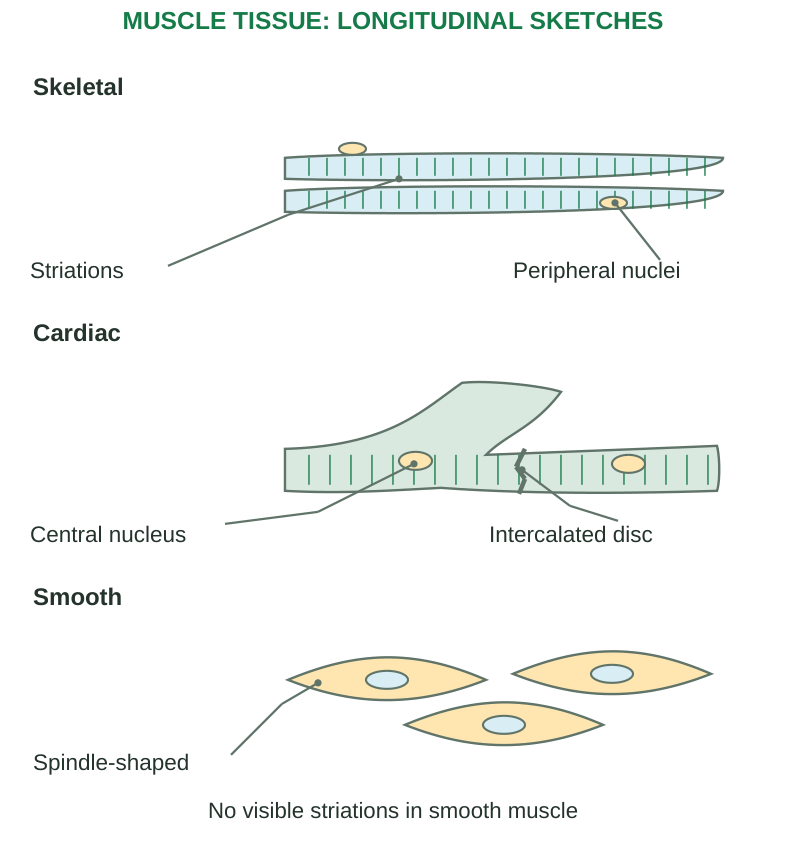
Diagram labels: Skeletal: striations and peripheral nuclei. Cardiac: central nucleus and intercalated disc. Smooth: spindle-shaped. No visible striations in smooth muscle.
| Feature | Skeletal | Cardiac | Smooth |
|---|---|---|---|
| Cell shape | Long cylindrical fibres | Shorter branching cells | Spindle-shaped cells |
| Striations | Present | Present | Not visible as sarcomeric bands |
| Nuclei | Many, usually peripheral | Usually one central, sometimes two | One central |
| Control | Mainly voluntary somatic control | Involuntary, intrinsically active with regulation | Involuntary with varied controls |
| Special feature | Organized sarcomeres | Intercalated discs | Dense bodies and different filament organization |
| Location | Muscles associated with skeleton and other selected sites | Myocardium | Walls of hollow organs and many vessels |
| Main function | Movement, posture and heat | Pumping blood | Moving contents and controlling diameter |
Striations
Repeated sarcomeric organization produces visible bands in skeletal and cardiac muscle. Smooth muscle contains actin and myosin but lacks the same regular sarcomeric appearance. “No striations” does not mean “no contractile proteins.”
Cardiac Intercalated Discs
Mechanical junctions link cells and gap junctions support coordinated electrical activity. Cardiac cells are separate cells joined functionally; the myocardium is not simply one giant multinucleated fibre.
Structure-Function Links
Long skeletal fibres generate directed force. Branching cardiac cells form coordinated networks. Smooth muscle can sustain contraction and regulate hollow-organ or vessel activity.
Exam trap: Voluntary does not mean every contraction requires conscious thought; skeletal reflexes also occur. Involuntary does not mean absence of regulation by nerves or other signals.
1.22 Nervous Tissue: Neurons and Glia
Nervous tissue receives, processes and transmits information. Neurons provide specialized signalling; glial cells support, protect, regulate and interact with neuronal function.
Neuron Structure
The cell body contains the nucleus and much metabolic machinery. Dendrites receive many inputs. The axon extends from an initial region and conducts signals toward terminals. Terminals communicate with target cells at synapses.
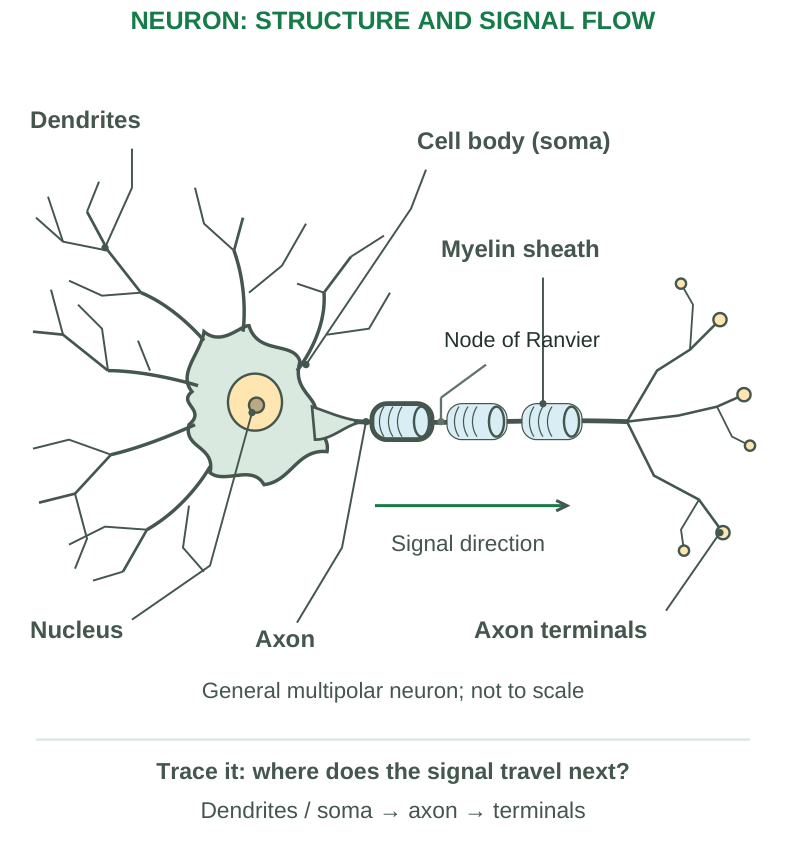
Diagram labels: Dendrites; cell body (soma); myelin sheath; nucleus; axon; axon terminals; signal direction; node of Ranvier. General multipolar neuron, not to scale. Trace it: where does the signal travel next? Dendrites/soma → axon → terminals.
Myelin
A myelin sheath surrounds segments of many axons, improving conduction properties. Nodes of Ranvier interrupt the sheath. Myelin is produced by oligodendrocytes in the CNS and Schwann cells in the PNS. Not every axon is myelinated.
Main Glial Groups
| Group | Important contribution |
|---|---|
| Astrocytes | Support extracellular conditions and participate in CNS functions/barrier relationships |
| Oligodendrocytes | CNS myelin formation |
| Microglia | CNS immune surveillance and responses |
| Ependymal cells | Line ventricles and central canal; specialized forms participate in CSF-related functions |
| Schwann cells | PNS axon support and myelin |
| Satellite cells | Support neuronal cell bodies in peripheral ganglia |
Locations and Functions
Nervous tissue forms brain, spinal cord, nerves and ganglia. Coordination involves sensory input, integration and output. Signals may influence muscle, glands or other neurons.
Renewal and Injury
Many mature neurons have very limited replacement capacity. Peripheral axonal regrowth may occur in suitable circumstances, but it is not guaranteed and differs from replacing the entire neuron. CNS injury and peripheral injury have different repair environments.
Study rule: Label the soma, nucleus, dendrites, axon, myelin and terminals. Do not label a dendritic branch as an axon or assume every neuron has the multipolar shape shown in a general sketch.
1.23 Cellular Injury: Meaning and Causes
Cell injury occurs when a harmful stimulus exceeds a cell’s ability to adapt or maintain normal function. Outcome depends on the nature, severity and duration of injury and the cell’s type, metabolic state and reserves. Mild, brief injury may be reversible; severe or persistent injury can cause death.
Major Causes
| Cause | Examples and mechanism |
|---|---|
| Oxygen deprivation | Hypoxia lowers oxygen availability; ischemia reduces blood supply, also reducing nutrients and waste removal |
| Physical agents | Trauma, extremes of temperature, radiation, electricity and pressure changes |
| Chemicals and drugs | Toxic substances or harmful drug/metabolite exposure; effect depends on dose and susceptibility |
| Infectious agents | Viruses, bacteria, fungi and parasites; direct injury or host responses |
| Immune reactions | Excessive or misdirected immune responses, including autoimmune injury |
| Genetic abnormalities | Altered proteins, enzymes or chromosomes may impair cell function |
| Nutritional imbalance | Deficiencies, excesses and imbalanced intake can damage tissues |
| Age-related changes | Accumulated damage and reduced repair capacity increase vulnerability |
Hypoxia Versus Ischemia
Hypoxia means inadequate oxygen at tissue level. Ischemia means inadequate blood flow. Ischemia usually produces hypoxia, but hypoxia can occur despite blood flow—for example, reduced oxygen carriage in severe anaemia.
Think in mechanisms: A cause is the initiating stimulus. Pathogenesis is the sequence linking that cause to functional and structural changes. Morphology describes the resulting appearance.
1.24 Membrane and Mitochondrial Damage
Membrane Injury
Damage may affect the plasma membrane, mitochondrial membranes or lysosomal membranes. Reduced ATP, calcium-dependent enzyme activity, reactive oxygen species and altered lipid metabolism contribute to membrane dysfunction.
- Plasma membrane failure disturbs ion gradients and permits loss of cellular contents.
- Mitochondrial membrane damage impairs energy production and can release proteins that activate apoptosis.
- Lysosomal membrane damage releases enzymes that digest cellular components.
Mitochondrial Injury
Mitochondria produce much cellular ATP by oxidative phosphorylation. Oxygen deprivation and other insults may reduce ATP production. Severe damage may open permeability pathways and prevent recovery of normal mitochondrial function.
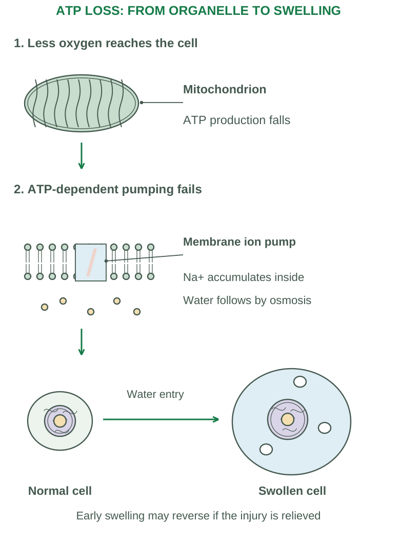
Diagram labels: Less oxygen reaches the cell: mitochondrion, ATP production falls. ATP-dependent pumping fails: membrane ion pump, Na+ accumulates inside, water follows by osmosis. Water entry; normal cell; swollen cell. Early swelling may reverse if the injury is relieved.
ATP Depletion: Linked Consequences
Na+/K+ pump failure causes intracellular sodium accumulation and water entry, producing swelling. Calcium regulation also becomes impaired. Greater reliance on anaerobic glycolysis depletes glycogen and promotes lactate accumulation and lower pH. Ribosome detachment from rough ER reduces protein synthesis.
Clinical Reasoning Example
An ischemic cell may first swell while retaining the possibility of recovery. Persistent ischemia can cause severe mitochondrial and membrane damage, making injury irreversible. Appearance alone should be interpreted with the tissue and injury context.
1.25 Ribosome, Nuclear, Calcium and Oxidant Injury
Ribosome Damage and Reduced Protein Synthesis
ATP depletion, swelling of the endoplasmic reticulum and ribosome detachment from rough ER impair translation. Reduced synthesis affects enzymes, structural proteins and proteins required for normal cell maintenance. A damaged cell may also accumulate unfolded proteins, activating stress responses.
Nuclear Damage
DNA injury can arise from radiation, reactive molecules and other insults. Cells may pause the cycle and activate repair. If damage cannot be repaired, apoptosis or loss of normal function may follow. Nuclear injury is not defined only by visible nuclear fragmentation.
Excess Intracellular Calcium
Calcium normally remains low in cytosol relative to extracellular fluid and intracellular stores. Injury may permit calcium entry or release from stores. Calcium can activate phospholipases, proteases, endonucleases and ATPases, worsening membrane, protein, DNA and energy disturbances.
Reactive Oxygen Species (ROS)
Examples include superoxide and hydrogen peroxide; the hydroxyl radical is highly reactive. Excess ROS can cause lipid peroxidation, protein modification and DNA damage. Antioxidant systems include glutathione, catalase and superoxide dismutase. ROS also participate in normal signalling; injury depends on imbalance.
| Target | Functional consequence |
|---|---|
| Membrane lipids | Loss of membrane integrity and gradients |
| Mitochondria | ATP depletion and death pathway activation |
| Ribosomes/ER | Reduced or abnormal protein production |
| Nucleus/DNA | Repair response, altered function or death |
Integrated answer: These mechanisms reinforce each other; do not present each damaged organelle as an entirely separate disease.
1.26 Reversible and Irreversible Injury
Reversible Injury
Early functional and structural changes may resolve if the stimulus is removed in time. Common features include cellular swelling, membrane blebs, mitochondrial swelling and ER dilation. Fatty change is important in some tissues, especially liver.
Cellular Swelling
Impaired energy-dependent ion regulation leads to sodium and water accumulation. Hydropic change describes pronounced swelling with clear cytoplasmic vacuoles, often reflecting dilated ER. Swelling is a morphological sign of injury, not a specific diagnosis or proof of cell death.
Irreversible Injury
Critical features include inability to restore mitochondrial function and severe membrane damage. Once essential integrity is lost, removing the original insult may no longer restore survival.
| Feature | Reversible injury | Irreversible injury/necrosis |
|---|---|---|
| Energy function | Reduced; recovery possible | Persistent major failure |
| Membrane | Blebs and altered transport | Severe disruption and leakage |
| Organelles | Swelling, ER changes | Destruction and enzyme release |
| Nucleus | Chromatin clumping may occur | Pyknosis, karyorrhexis or karyolysis |
Nuclear Changes in Cell Death
- Pyknosis: Nuclear shrinkage with increased staining.
- Karyorrhexis: Fragmentation of the condensed nucleus.
- Karyolysis: Fading/dissolution of nuclear staining through DNA degradation.
These describe necrotic nuclear morphology; their exact visibility depends on timing and tissue.
Injury Versus Adaptation
Adaptation changes cell size, number or phenotype to cope with demands. It may maintain survival but also reduce reserve. Injury occurs when the stress exceeds that capacity; adaptation and injury may coexist.
1.27 Atrophy, Hypertrophy and Hyperplasia
Adaptation is a change that helps cells respond to altered demand or stress. The response depends on the tissue’s ability to grow, divide and change phenotype.
| Change | Definition | Typical example |
|---|---|---|
| Atrophy | Reduced cell size, often with reduced tissue mass and sometimes fewer cells | Muscle wasting after immobilization |
| Hypertrophy | Increased cell size and organ size | Enlarged skeletal muscle with training; cardiac enlargement with pressure load |
| Hyperplasia | Increased cell number in a tissue capable of proliferation | Hormone-related breast gland growth; compensatory liver growth |
Atrophy
Reduced workload, loss of nerve supply, reduced blood supply, poor nutrition, reduced endocrine stimulation and aging can contribute. Protein synthesis falls, while protein breakdown and autophagy may increase. Atrophy is a reduction of previously developed tissue; hypoplasia refers to underdevelopment.
Hypertrophy
Greater synthesis of structural proteins and organelles enlarges cells. It is especially important in tissues with limited capacity for cell division, such as mature cardiac muscle. Physiological enlargement can meet increased demand; sustained pathological load can eventually cause dysfunction.
Hyperplasia
Growth signals stimulate proliferation of mature cells or tissue stem cells. It may be physiological, compensatory or pathological. Hormonal stimulation can cause pathological hyperplasia. Hyperplasia remains responsive to growth controls and is not synonymous with neoplasia.
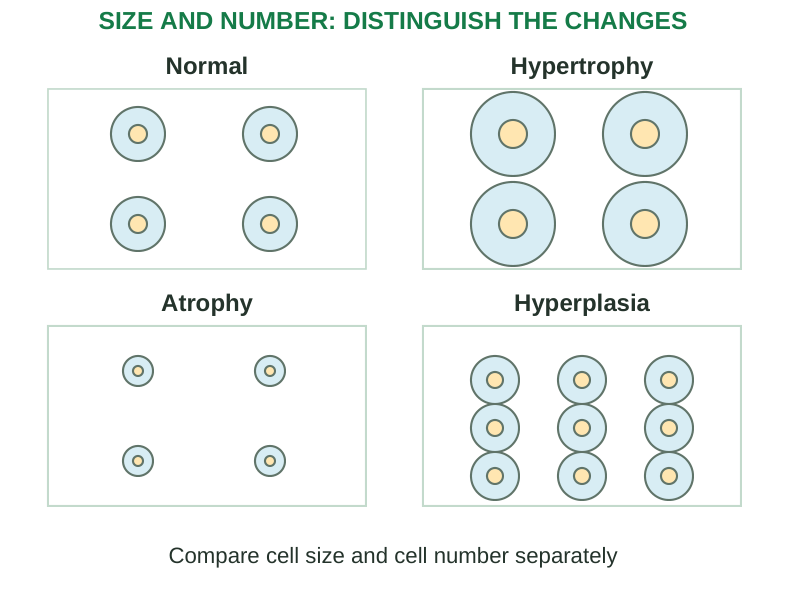
Diagram labels: Normal; hypertrophy; atrophy; hyperplasia. Compare cell size and cell number separately.
Compare Carefully
Size increases in hypertrophy; number increases in hyperplasia. Both may occur together, as in the pregnant uterus. A larger organ does not tell you which process occurred without cellular evidence.
1.28 Metaplasia and Dysplasia
Metaplasia
Metaplasia is replacement of one differentiated cell type by another better suited to a persistent stress. It often results from altered differentiation of stem or precursor cells rather than a mature cell simply changing shape.
Example: chronic airway irritation may replace normal ciliated columnar epithelium with stratified squamous epithelium. The replacement tolerates irritation better but loses important mucociliary functions. Removing the stimulus may allow reversal.
Dysplasia
Dysplasia is disordered growth and maturation, often with cellular atypia and disturbed tissue architecture. Cells may vary in size and shape, have enlarged dark nuclei and show abnormal proliferation or loss of normal organization.
Dysplasia is commonly discussed with adaptive/morphological changes in introductory syllabi, but it is not a protective adaptation in the same sense as hypertrophy or metaplasia. It is not automatically invasive cancer. Some dysplastic lesions regress; others progress depending on context and grade.
| Point | Metaplasia | Dysplasia |
|---|---|---|
| Main change | Differentiated cell type replaced | Growth and maturation become disordered |
| Organization | May remain orderly | Cellular/architectural atypia |
| Response to stress | May improve tolerance at functional cost | Indicates abnormal development/growth |
| Cancer relationship | Persistent causes may increase risk | Some lesions are precancerous; not all progress |
Do Not Confuse These Terms
Neoplasia is abnormal new growth that becomes relatively autonomous. Invasion means extension through normal tissue boundaries. Neither term can be substituted for metaplasia, hyperplasia or every instance of dysplasia.
Memory line: Atrophy: smaller; hypertrophy: bigger cells; hyperplasia: more cells; metaplasia: different mature cell type; dysplasia: disorder.
1.29 Intracellular Accumulations
Substances may accumulate because normal metabolism, transport, folding or degradation is disturbed. The accumulated material may be a normal constituent present in excess, an abnormal endogenous product or a foreign substance. Consequences range from little effect to substantial injury.
| Material | Example and significance |
|---|---|
| Lipids | Triglyceride accumulation in hepatocytes produces fatty change; cholesterol may accumulate in some lesions |
| Proteins | Excess reabsorbed protein in renal tubules or abnormal/misfolded proteins in cells |
| Glycogen | Excess accumulation in some metabolic conditions; appears as clear cytoplasmic material with appropriate preparation |
| Lipofuscin | Residual pigment associated with accumulated cellular wear; often seen with aging |
| Hemosiderin | Iron-storage pigment; local or systemic excess may reflect iron accumulation |
| Bilirubin | Bile pigment that may accumulate in cholestatic states |
| Exogenous pigment | Carbon particles can be taken up by macrophages |
Fatty Change
Steatosis means abnormal triglyceride accumulation in parenchymal cells. Altered fatty acid delivery, oxidation, lipid synthesis or export may contribute. The liver is a common site because of its role in lipid metabolism. Fatty change and obesity are different concepts.
Intracellular Versus Extracellular
Intracellular accumulation is material within cells. An extracellular deposit is outside cells. Calcification and other deposits may be discussed in wider pathology, but they should not all be described as intracellular accumulation.
Interpretation
A stain or appearance suggests a substance; identification may require appropriate tests. An accumulation does not automatically prove irreversible injury, and pigment itself may be less harmful than the underlying disorder.
1.30 Cell Death: Necrosis and Apoptosis
Necrosis describes cell death associated with severe injury, loss of membrane integrity and tissue reaction. Apoptosis is regulated cell death involving controlled dismantling, typically with little inflammation when fragments are promptly cleared.
| Feature | Necrosis | Apoptosis |
|---|---|---|
| Common setting | Severe pathological injury | Physiological turnover or pathological signals |
| Cell appearance | Swelling and breakdown | Shrinkage and condensation |
| Membrane | Integrity lost | Remains intact during fragmentation; later cleared |
| Contents | Leak into tissue | Packaged in apoptotic bodies |
| Inflammation | Usually present | Usually minimal with efficient clearance |
| Distribution | Often groups of cells | Often individual cells |
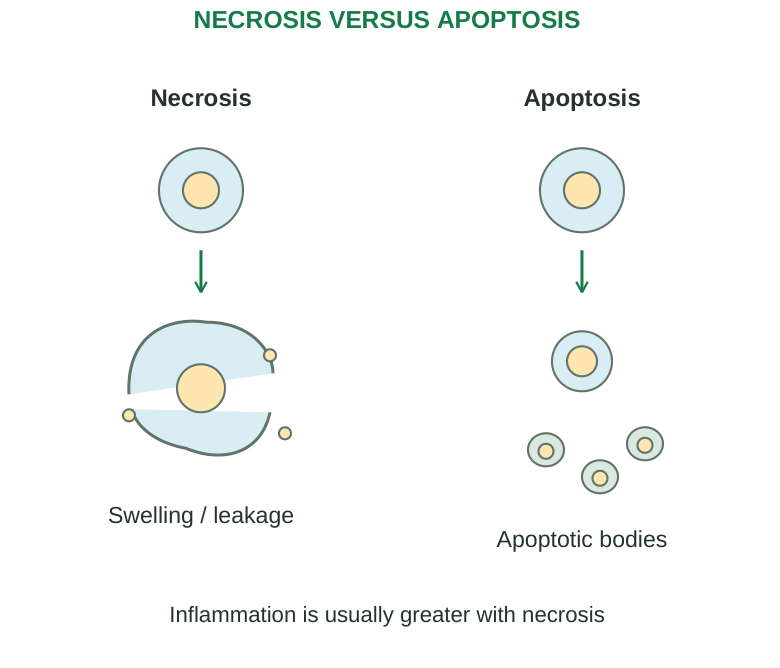
Diagram labels: Necrosis: swelling/leakage. Apoptosis: apoptotic bodies. Inflammation is usually greater with necrosis.
Apoptotic Pathways
The intrinsic pathway responds to internal stress, including unrepaired DNA damage, through mitochondrial signalling. The extrinsic pathway begins at certain cell-surface death receptors. Both can activate caspases that dismantle the cell. Apoptosis supports development, immune regulation and removal of unnecessary or damaged cells.
Main Necrosis Patterns
- Coagulative: Tissue outline initially preserved; common in ischemic injury of solid organs except brain.
- Liquefactive: Digestion converts tissue to liquid; seen in abscesses and many brain infarcts.
- Caseous: Soft, cheese-like debris; classically associated with tuberculosis.
- Fat necrosis: Destruction of fat, for example with pancreatic enzyme release; may produce calcium soaps.
- Fibrinoid: Characteristic vessel-wall injury associated with some immune or severe vascular conditions.
Gangrene is a clinical term, commonly applied to ischemic tissue necrosis; infection may add liquefaction in wet gangrene. It is not a separate basic microscopic pattern.
1.31 Common Medical Terminology
| Term | Meaning |
|---|---|
| Etiology | Cause or causal factors of a condition |
| Pathogenesis | Mechanism and development of disease |
| Morphology | Structural appearance of cells/tissues |
| Lesion | Localized structural or functional abnormality |
| Sign | Objective finding detected by examination/testing |
| Symptom | Subjective experience reported by a person |
| Syndrome | Recognizable group of associated findings |
| Diagnosis | Identification of a condition from evidence |
| Prognosis | Expected course or outcome |
| Acute | Rapid onset or relatively short course |
| Chronic | Persistent or prolonged course |
| Idiopathic | Cause not established |
| Iatrogenic | Caused by medical intervention |
| Inflammation | Tissue response to injury/infection involving immune and vascular changes |
| Oedema | Excess fluid in interstitial spaces or body cavities |
| Hypoxia | Inadequate tissue oxygen availability |
| Ischemia | Reduced tissue blood supply |
| Infarction | Area of ischemic necrosis |
| Degeneration | Deteriorative changes in cells or tissues |
| Adaptation | Adjustment to altered demand or stress |
| Regeneration | Replacement through growth of appropriate tissue cells |
| Fibrosis | Excess deposition of fibrous connective tissue |
Word Parts
- Hyper-: Above/excessive.
- Hypo-: Below/reduced.
- Intra-: Within.
- Extra-: Outside.
- Peri-: Around.
- -itis: Commonly indicates inflammation.
- -ectomy: Surgical removal.
- -algia: Pain.
- -emia: A blood-related condition.
Precision matters: A symptom is not a diagnosis. Inflammation does not always mean infection. Acute/chronic describe a course and do not by themselves establish severity.
1.32 Worked Cases and Rapid Revision
Case 1: A Red Blood Cell in Dilute Fluid
If the surrounding solution is sufficiently hypotonic, water enters by osmosis. The red cell swells and may undergo haemolysis. A hypertonic solution instead causes water loss and shrinkage. Tonicity depends mainly on effectively non-penetrating solutes.
Case 2: Five Sodium-Potassium Pump Cycles
Each cycle uses one ATP, exports three Na+ and imports two K+.
Five cycles:
- 5 ATP used.
- 15 Na+ out.
- 10 K+ in.
This is a stoichiometric calculation; it does not specify total cellular pumping rate.
Case 3: Chromosome Counting
A human somatic cell has 46 chromosomes before DNA replication. After S phase it still has 46 chromosomes, now consisting of 92 chromatids. Following normal mitosis and cytokinesis, each daughter cell has 46 chromosomes. Chromosomes are counted by centromeres.
Case 4: Identify Signalling
- A membrane-bound ligand contacting a neighbouring cell: contact-dependent.
- A local mediator reaching nearby cells: paracrine.
- A neuron releasing transmitter across a synapse: synaptic.
- A hormone carried through blood to distant targets: endocrine.
Case 5: Identify Adaptation
- Muscle enlargement from increased workload: hypertrophy.
- Immobilized muscle becoming smaller: atrophy.
- Increased gland-cell number under hormones: hyperplasia.
- Airway epithelial replacement under persistent irritation: metaplasia.
- Disordered atypical maturation: dysplasia.
Case 6: Connect Oxygen Loss to Swelling
Ischemia reduces oxygen delivery → oxidative phosphorylation falls → ATP falls → Na+/K+ pumping fails → sodium and water accumulate → swelling
Persistent injury may progress to membrane failure and death.
One-Minute Tissue Revision
- Epithelium: Covers, lines and secretes.
- Connective: Supports, binds and transports.
- Muscle: Contracts.
- Nervous: Communicates and coordinates.
Add structure, location and a specific function to every examination answer.
1.33 Examination Practice
Long-Answer Practice
- 1. Define homeostasis. Describe a feedback system with components and explain negative and positive feedback using examples.
- 2. Draw a labelled generalized cell and explain the functions of major organelles.
- 3. Classify membrane transport. Compare passive and active mechanisms, with examples.
- 4. Classify epithelial and connective tissues. Explain their structure, locations and functions.
- 5. Describe causes and mechanisms of cell injury, including membrane, mitochondrial, ribosomal and nuclear changes.
- 6. Compare adaptations, reversible injury, necrosis and apoptosis with examples.
Short-Answer Practice
- Anatomical position, planes and directional terms.
- Mitosis versus meiosis; cell-cycle checkpoints.
- Tight junction, desmosome and gap junction.
- Four forms of intercellular signalling.
- Skeletal, cardiac and smooth muscle.
- Neuron, myelin and main glial groups.
- Atrophy versus hypertrophy versus hyperplasia.
- Metaplasia versus dysplasia; intracellular accumulations.
Build a Useful Answer
Start with a precise definition. Classify if required. Explain the mechanism or structure, then add locations/examples and a labelled figure. End with the relevant function or consequence. Match the detail to the marks available.
Test Yourself: Answers Below
- 1. Which junction permits small molecules/ions to pass between adjacent cells?
- 2. Which membrane transport mechanism directly uses ATP to move three Na+ out and two K+ in?
- 3. Which epithelial type permits distension in the urinary tract?
- 4. What is replacement of one differentiated cell type by another called?
- 5. Name nuclear shrinkage, fragmentation and fading in necrosis.
- 6. A local chemical acts on neighbouring cells. Which signalling form is this?
Answers:
- 1. Gap junction.
- 2. Na+/K+-ATPase.
- 3. Transitional epithelium/urothelium.
- 4. Metaplasia.
- 5. Pyknosis, karyorrhexis, karyolysis.
- 6. Paracrine.
Worked Distinction
Question: A larger tissue contains enlarged cells but no increase in number. Name the change and distinguish it from hyperplasia.
Answer: Hypertrophy; cells become larger. Hyperplasia increases cell number in a tissue capable of proliferation. Both can coexist, but this description specifies hypertrophy.
Final Revision Checklist
- Define anatomy, physiology and pathophysiology.
- Trace structural levels and recall body systems/life processes.
- Use anatomical terms and feedback components correctly.
- Label the cell, membrane, tissue and neuron figures.
- Compare transport, division, junctions and signalling.
- Explain all four tissue classes with locations/functions.
- Connect cell-injury causes to mechanisms and morphology.
- Distinguish all five named cellular changes and both death processes.
- Revise terminology and practise diagrams without crossed labels.
Unit I Complete
Revise every syllabus heading. Practise one labelled sketch and one mechanism-based answer in each study session.