
Course Code: BP104T
Unit II Introduction
Integumentary System, Wound Healing, Skeletal System and Joints. This unit connects the body’s protective covering with its supporting framework and movement machinery. It explains normal structure first, then the mechanisms behind important skin, bone and joint disorders.
The skin limits water loss and provides protection and sensation. Bones support the body, store minerals and provide levers. Skeletal muscles generate force, while joints determine where and how movement occurs. Injury and disease alter these relationships.
Topics Covered – Complete Syllabus Map
| Required topic | Sections |
|---|---|
| Skin structure and functions | 2.1–2.4 |
| Psoriasis and dermatitis | 2.5–2.6 |
| Pathophysiology of leprosy | 2.7 |
| Principles of wound healing | 2.8–2.10 |
| Skeletal divisions and types of bones | 2.11–2.12 |
| Bone structure and functional basis | 2.13–2.14 |
| Axial skeletal bones and functions | 2.15 |
| Appendicular bones and functions | 2.16 |
| Organization of skeletal muscle | 2.17–2.18 |
| Neuromuscular junction | 2.19 |
| Physiology of contraction | 2.20–2.22 |
| Structural/functional joint classification | 2.23–2.24 |
| Rheumatoid arthritis | 2.25 |
| Osteoporosis and gout | 2.26–2.27 |
| Revision, cases and exam practice | 2.28–2.29 |
Learning Outcomes
- Identify skin layers and explain how each supports function.
- Distinguish inflammatory skin disease from infection.
- Explain wound repair through overlapping stages.
- Classify bones and locate major axial and appendicular groups.
- Trace muscle organization down to myofilaments.
- Explain neuromuscular transmission, calcium activation and cross-bridge cycling.
- Compare joint classifications and movements.
- Explain mechanisms of the three required bone/joint diseases.
Essential Vocabulary
| Term | Core meaning |
|---|---|
| Epidermis | Outer epithelial skin layer |
| Dermis | Supporting connective tissue of skin |
| Granulation tissue | New vascular connective tissue in repair |
| Osteoblast | Bone-forming cell |
| Osteoclast | Bone-resorbing cell |
| Sarcomere | Contractile unit between Z discs |
| Motor unit | One motor neuron and its muscle fibres |
| Synovium | Specialized lining of a synovial joint |
| Pannus | Invasive inflamed tissue in RA |
| Tophi | Deposits containing urate crystals |
Study Route
Structure → function → disturbance → consequences.
For every disorder, learn the defining mechanism before memorizing features. For every sketch, identify the structure and explain one function of each major label.
Central question: How does a change in a barrier, a supporting tissue or a signalling mechanism alter protection, repair or movement?
2.1 Integumentary System and Skin Layers
The integumentary system includes skin and its associated hair, nails and glands. Skin consists of an epidermis and a dermis. The hypodermis lies beneath them and connects skin to deeper structures; it is not usually counted as skin proper.
Layers at a Glance
| Region | Tissue and main importance |
|---|---|
| Epidermis | Keratinized stratified squamous epithelium; external barrier |
| Dermis | Connective tissue; strength, elasticity, vessels, nerves and appendages |
| Hypodermis | Loose connective tissue and variable fat; anchorage, cushioning and insulation |
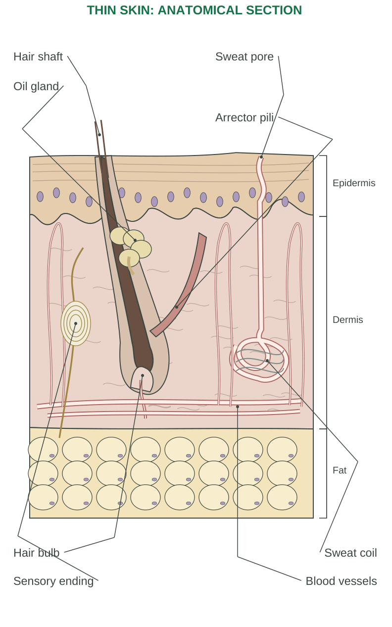
Diagram labels: Hair shaft; sweat pore; oil gland; arrector pili; epidermis; dermis; fat; hair bulb; sweat coil; sensory ending; blood vessels.
Avascular Versus Vascular
The epidermis has no blood vessels. Nutrients reach its living cells by diffusion from the dermis. The dermis contains vessels and supports the epidermis. Damage extending into dermis can therefore produce bleeding; superficial loss of only epidermal layers may not.
Thick and Thin Skin
Thick skin is found on palms and soles. It has a thick epidermis, a distinct stratum lucidum and no hair follicles or sebaceous glands. Thin skin covers most of the body and generally has hair follicles. “Thick skin” in histology refers mainly to the epidermis, not simply to the total depth of all tissues.
Why the Arrangement Matters
An outer epithelial barrier resists abrasion and limits water loss. Deeper collagen-rich tissue provides strength, while vessels and nerves permit regulation and sensation. A diagram should show the hypodermis below the dermis, rather than incorrectly placing fat within the epidermis.
2.2 Epidermis: Strata and Cell Types
From deep to superficial, the epidermal strata are basale, spinosum, granulosum, lucidum in thick skin, and corneum.
| Stratum | Main feature and contribution |
|---|---|
| Basale | Deep basal cells attached to basement membrane; proliferative population renews epidermis |
| Spinosum | Cells connected by strong junctions; contributes mechanical integrity |
| Granulosum | Keratinization-related granules and lipid barrier formation |
| Lucidum | Clear layer recognizable in thick skin |
| Corneum | Flattened dead corneocytes with keratin, embedded in a lipid-rich barrier |
Major Cell Types
Keratinocytes form most of the epidermis and develop the protective surface. Melanocytes, located chiefly in the basal region, produce melanin and transfer pigment to keratinocytes. Epidermal dendritic/Langerhans cells participate in immune surveillance. Merkel cells associate with sensory nerve endings and contribute to touch sensation.
Renewal and Pigmentation
Basal proliferation, differentiation, upward movement and shedding maintain the epidermis. Renewal rates vary with body site and conditions. Pigmentation depends on melanin production, distribution and other factors, not simply the number of melanocytes.
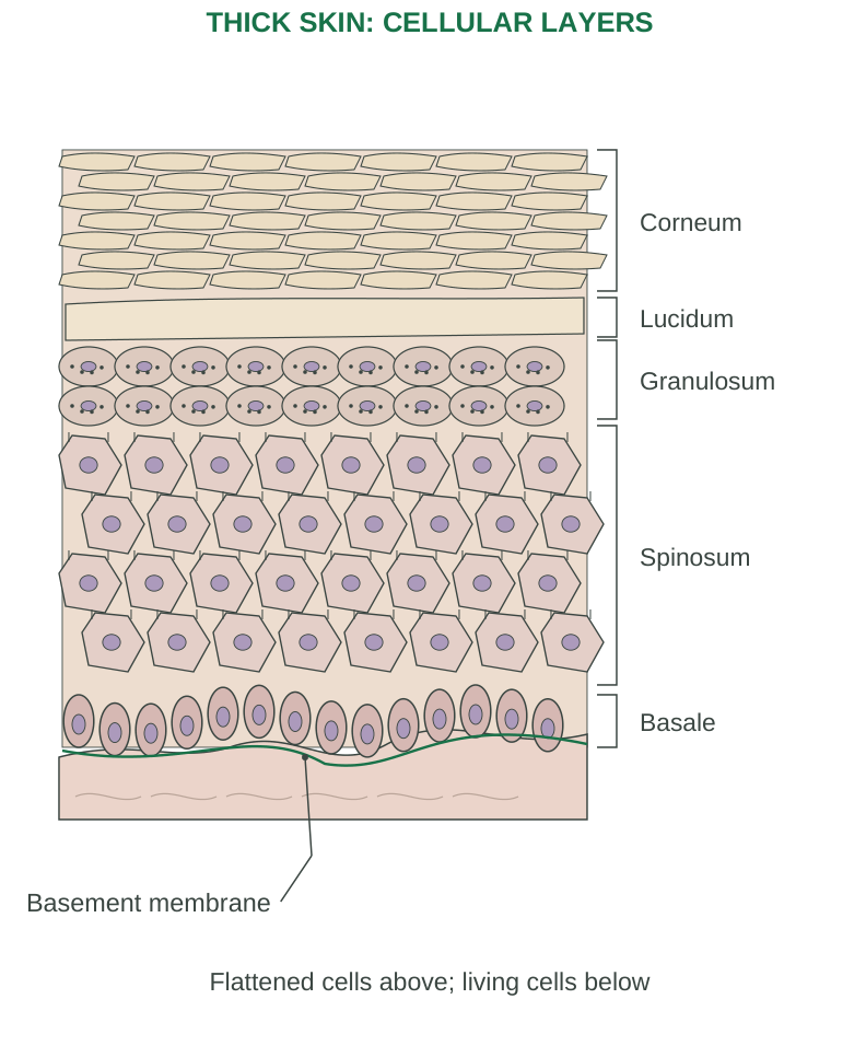
Diagram labels: Corneum; lucidum; granulosum; spinosum; basale; basement membrane. Flattened cells above; living cells below.
Barrier Concept
The stratum corneum can be understood as protein-rich cells held in a lipid-rich extracellular environment. Barrier damage increases water loss and permits greater penetration of irritants. This connects normal structure to dermatitis and wound care.
Memory aid: B–S–G–L–C: Basale → Spinosum → Granulosum → Lucidum → Corneum. Add the qualification: lucidum is characteristic of thick skin.
2.3 Dermis, Hypodermis and Appendages
Dermis
The superficial papillary region contains relatively loose connective tissue and forms dermal papillae. The deeper reticular region contains dense irregular connective tissue rich in collagen and elastic fibres. Together they support vessels, nerves, glands and hair-associated structures.
Hair and Associated Structures
A hair shaft emerges through the surface; its root lies within a follicle. The bulb contains growth-related cells, supported by a dermal papilla. Sebaceous glands commonly empty into follicles. The arrector pili is smooth muscle that can raise the hair and produce goosebumps.
Skin Glands
| Gland | Secretion and function |
|---|---|
| Eccrine sweat | Mainly watery secretion to surface through a duct; evaporation contributes to cooling |
| Apocrine sweat | Mainly in selected regions such as axilla; secretion into follicles; bacterial processing contributes to odour |
| Sebaceous | Sebum, usually into follicles; lubrication and surface protection |
Nails
Nails are keratinized plates. The nail matrix produces the plate, while the underlying nail bed supports it. Their hard surface protects distal digits and assists fine manipulation. Nails do not generate force independently of the surrounding tissues.
Hypodermis
Adipose and loose connective tissues permit mobility over deeper structures and provide cushioning, insulation and energy storage. The amount and distribution of fat vary greatly between body regions and individuals.
Sensory Structures
Free nerve endings and specialized receptors contribute to pain, temperature, touch and pressure. Receptor location and specialization differ. A labelled skin sketch should show neural structures in the dermis or deeper regions, rather than presenting the outer dead corneum as a functioning nerve layer.
2.4 Functions of Skin and Thermoregulation
| Function | Structural or physiological basis |
|---|---|
| Protection | Keratinized epithelium, lipids, immune defences and pigmentation |
| Water balance | Surface barrier limits excessive water loss |
| Sensation | Sensory nerves and specialized receptors |
| Temperature regulation | Sweating and altered skin blood flow |
| Vitamin D formation | UV-dependent conversion of a skin precursor; later activation involves liver and kidney |
| Immune contribution | Barrier and local immune-cell responses |
| Limited excretion | Sweat contains water, salts and small amounts of other substances |
Heat Balance
When heat loss must increase, sweating and skin vasodilation help transfer heat to the environment. Evaporation removes heat; sweat that drips away without evaporating provides less cooling. High humidity can reduce evaporative efficiency. Vasoconstriction helps conserve heat in cold conditions.
Barrier Disruption: An Integrated Example
A large skin injury can disturb fluid balance, permit infection and alter temperature regulation. These effects follow from loss of several normal skin functions, not only from discomfort at the injured area.
Vitamin D Qualification
Skin initiates vitamin D production, but does not by itself perform all steps required to produce the active hormone. Sunlight exposure is not a guarantee of adequate vitamin D in every individual.
Excretion Qualification
Skin is not the principal organ for removal of metabolic wastes. Kidneys and other organs remain central. Describing sweat as a way to remove “all toxins” is inaccurate.
Answer pattern: Name a function, identify the supporting structure, and give a mechanism. Example: temperature control—eccrine gland—sweat evaporation removes heat.
2.5 Psoriasis: Pathophysiological Basis
Psoriasis is an immune-mediated inflammatory skin disease associated with altered keratinocyte proliferation and differentiation. It is not a contagious fungal infection.
Mechanism
Genetic susceptibility and environmental influences contribute to immune activation. T-cell-related cytokine signalling, including important IL-23/IL-17 pathways, sustains inflammation. Epidermal turnover and maturation become abnormal, producing thickened skin and surface scaling.
Typical Features
Plaque psoriasis commonly causes well-demarcated, raised scaly lesions. Extensor regions, scalp and other sites may be involved. Appearance varies with skin tone. Nail pitting or separation may occur, and some people develop psoriatic arthritis.
Morphological Link
Acanthosis means epidermal thickening. Parakeratosis means retention of nuclei in cells of the cornified layer. Inflammation and altered vascular patterns contribute to visible changes. The pattern is more informative than describing every lesion simply as “red skin.”
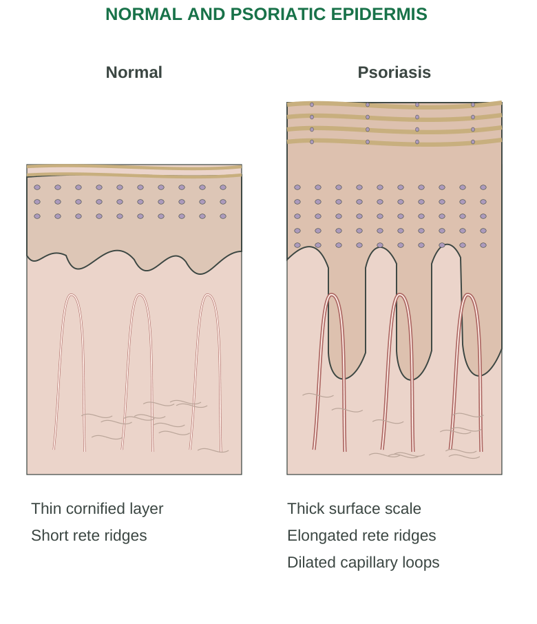
Diagram labels: Normal and psoriatic epidermis. Normal: thin cornified layer and short rete ridges. Psoriasis: thick surface scale, elongated rete ridges and dilated capillary loops.
Triggers and Course
The disease often fluctuates. Skin injury can provoke lesions at injured sites in susceptible people, called the Koebner phenomenon. Infections, stress and some medicines may influence activity. A trigger does not replace the underlying immune mechanism.
Study Distinction
The visible scale is the consequence of altered epidermal biology. It does not mean that the primary problem is ordinary dirt or a lack of hygiene. Psoriatic arthritis and rheumatoid arthritis are distinct disorders.
2.6 Dermatitis: Barrier and Inflammatory Changes
Dermatitis means inflammation of the skin. The term includes several conditions and does not identify one universal cause.
| Type | Main underlying mechanism |
|---|---|
| Atopic dermatitis | Barrier dysfunction, immune dysregulation and environmental/genetic interactions |
| Irritant contact dermatitis | Direct injury from an irritant; prior sensitization is unnecessary |
| Allergic contact dermatitis | Delayed cell-mediated hypersensitivity after sensitization to a contact allergen |
Atopic Dermatitis
Reduced barrier function increases water loss and permits entry of irritants and other environmental substances. Inflammation further disturbs the barrier. Itching leads to scratching, producing an itch–scratch cycle that worsens damage. Not every person has the same genetic defect or trigger.
Contact Dermatitis
An irritant can damage skin directly, depending on strength and exposure. An allergen can trigger an immune response in a previously sensitized person. Nickel is a common example used to explain allergic contact disease. Irritant and allergic mechanisms may coexist.
Features and Consequences
Itching, dryness, erythema or colour change, scaling and sometimes vesicles or oozing can occur. Repeated rubbing may cause lichenification, meaning thickened skin with accentuated markings. Barrier disruption increases susceptibility to secondary infection.
Comparison with Psoriasis
Psoriasis often has sharply demarcated plaques and prominent scale; dermatitis often emphasizes itch and barrier disruption. Clinical overlap exists, so one visible feature alone does not establish the diagnosis.
Do not equate these: Dermatitis is not automatically contagious, caused by food allergy or equivalent to a bacterial infection. State the particular type and mechanism.
2.7 Leprosy: Skin–Nerve Pathophysiology
Leprosy, also called Hansen disease, is a chronic infection caused mainly by Mycobacterium leprae. It particularly affects skin and peripheral nerves. The organism favours relatively cooler tissues; host immune responses strongly influence the disease pattern.
Pathogenesis
The bacillus can infect macrophages and Schwann-cell-related nerve environments. Direct infection and immune inflammation contribute to peripheral nerve dysfunction. Loss of protective sensation permits repeated unnoticed trauma, while autonomic and motor dysfunction can further damage tissues.
Spectrum of Disease
Stronger cell-mediated immunity tends to limit organisms and produce more localized disease. Poorer containment permits a greater bacillary burden and more extensive involvement. Tuberculoid, borderline and lepromatous descriptions express an immunological spectrum; operational paucibacillary/multibacillary classifications serve a different purpose.
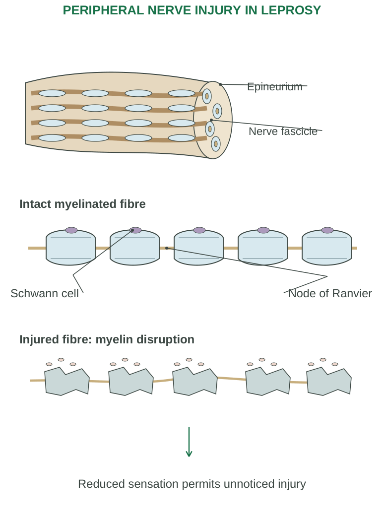
Diagram labels: Epineurium; nerve fascicle; intact myelinated fibre; Schwann cell; node of Ranvier. Injured fibre: myelin disruption. Reduced sensation permits unnoticed injury.
Key Manifestations
A pale or reddish patch with definite sensory loss, enlarged peripheral nerves with relevant sensory/motor deficits, and demonstration of bacilli are important diagnostic features. Weakness, deformity and injuries can follow nerve damage. Not every light-coloured patch is leprosy.
Important Principles
It is treatable and should not be interpreted as a hereditary curse or a result of casual brief contact. Early recognition limits nerve-related disability. Nerve reactions can require prompt evaluation even when infection treatment is underway.
Examination Chain
Skin/peripheral nerve infection → nerve dysfunction → sensory loss or weakness → repeated injury and disability
Separate primary disease from the secondary damage caused by loss of protection.
2.8 Wound Healing: Regeneration and Repair
Wound healing restores continuity after tissue injury. It combines regeneration of surviving appropriate cells with repair through extracellular matrix and scar formation. The balance depends on tissue type, damage depth and preservation of supporting structure.
What a Wound Disturbs
Skin injury disrupts the barrier. Deeper injury also damages vessels, connective tissue and sometimes appendages or nerves. Healing must control bleeding, remove damaged material, cover the surface and rebuild mechanical support.
Four Overlapping Phases
| Phase | Dominant events |
|---|---|
| Haemostasis | Vessel responses, platelet plug and fibrin clot |
| Inflammation | Leukocyte recruitment and removal of microbes/debris |
| Proliferation | New vessels, fibroblasts, matrix and epithelial coverage |
| Remodelling | Matrix reorganization and maturation of scar |
Overlap, Not Isolated Boxes
One phase does not completely finish before the next begins. Timings vary with wound size, tissue perfusion, infection and host factors. Memorizing one fixed day for every transition gives a misleading picture.
Cells and Matrix
Platelets help establish a clot and supply mediators. Neutrophils and macrophages contribute to defence and clearance. Fibroblasts produce matrix; endothelial cells form new vessels. Epithelial cells migrate and proliferate to restore surface coverage. Myofibroblasts contribute to wound contraction.
A Useful Distinction
Granulation tissue means vascular new repair tissue. A granuloma is a particular pattern of chronic inflammation. The similar names do not mean the same structure.
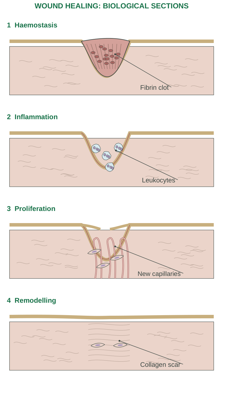
Diagram labels: Haemostasis: fibrin clot. Inflammation: leukocytes. Proliferation: new capillaries. Remodelling: collagen scar.
2.9 Healing Phases: Mechanisms and Outcome
Haemostasis
Vascular responses and platelets limit blood loss. Coagulation produces a fibrin network that stabilizes the clot. This provisional matrix also helps organize subsequent repair. A clot is not yet a mature scar.
Inflammatory Phase
Inflammatory cells remove damaged material and help control microbial contamination. Macrophages also coordinate later repair through signalling. Inflammation is necessary in controlled form; persistent excessive inflammation can delay closure.
Proliferative Phase
Angiogenesis provides new microvessels. Fibroblasts deposit extracellular matrix, initially rich in less mature collagen organization. Granulation tissue fills the defect. Keratinocytes advance across a suitable surface to restore epithelial continuity. Myofibroblast contraction reduces the area of some wounds.
Remodelling Phase
Collagen is reorganized, cross-linking increases and the vascularity/cellularity of the scar generally decline. Tensile strength improves over time, but healed scar tissue generally does not regain all properties of the original unwounded tissue. Hair follicles and other appendages may not be restored in a deep scar.
Structure Explains Outcome
A superficial epithelial defect can heal largely by epithelial regeneration if the supporting environment remains suitable. Deep loss of dermis usually requires more connective-tissue repair, increasing scar formation. Scar strength, surface appearance and restoration of specialized function are different outcomes.
Trace the sketch: Identify clot, inflammatory cells, new capillaries, fibroblasts, migrating epithelium and reorganized collagen. Explain how each changes between stages rather than drawing the same wound four times.
2.10 Healing Intention and Influencing Factors
| Type | Wound relationship and expected repair |
|---|---|
| Primary intention | Closely approximated edges, relatively little tissue loss; less granulation tissue and contraction |
| Secondary intention | Larger open defect; more granulation tissue, contraction and scar |
| Delayed primary closure | Wound initially managed open, then closed when appropriate |
Local Influences
Infection, foreign material, poor blood supply, repeated trauma, wound tension and extensive tissue loss can delay healing. Appropriate moisture supports epithelial repair, but excessive maceration damages surrounding skin. Oxygen delivery depends on perfusion, not only on oxygen being present in the air.
Systemic Influences
Poor nutrition, inadequate protein or relevant micronutrients, diabetes-related abnormalities, vascular disease, smoking and some medicines can impair repair. Age can influence reserve, but age alone does not determine healing outcome.
Complications
- Dehiscence: Separation of previously approximated wound edges.
- Ulceration: Persistent loss of surface tissue.
- Hypertrophic scar: Excessive scar within the original wound boundary.
- Keloid: Scar extends beyond that boundary.
- Contracture: Excessive contraction restricts movement, especially near joints.
Case Reasoning
A clean shallow cut and a large infected tissue defect need not heal at the same speed or by the same balance of regeneration and scar formation. Explain differences through tissue loss, perfusion, inflammation and edge relationship.
Practical Learning Rule
Avoid describing healing only as “new skin appears.” Include the vascular, cellular and matrix events underneath the visible surface.
2.11 Skeletal System: Divisions and Functions
The skeleton provides support, protection and a framework for movement. The typical adult count is 206 bones, although normal variation occurs. Separate bones in children can fuse during development.
| Division | Usual adult count | Components |
|---|---|---|
| Axial skeleton | 80 | Skull, auditory ossicles, hyoid, vertebral column and thoracic cage |
| Appendicular skeleton | 126 | Pectoral girdles, upper limbs, pelvic girdle and lower limbs |
Core Functions
- Support: Establishes body framework and attachment sites.
- Protection: Skull protects brain; thoracic cage protects major thoracic organs.
- Movement: Bones act as levers moved by muscles across joints.
- Mineral reservoir: Bone participates in calcium and phosphate homeostasis.
- Haematopoiesis: Red marrow produces blood cells.
- Energy storage: Yellow marrow contains substantial adipose tissue.
Bone Is Living Tissue
Blood supply, cells and ongoing remodelling distinguish bone from an inert mineral rod. Its strength comes from both organic matrix and mineral. A change in either component can disturb mechanical performance.
Bone, Cartilage and Ligament
Bone is mineralized connective tissue. Cartilage supports and cushions selected regions but is generally avascular. Ligaments connect bones and stabilize relationships; tendons connect muscle to its attachment, commonly bone.
Functional Example
The femur provides a strong lower-limb lever; muscle-generated force moves it through joints. Bone alone cannot actively contract. Movement requires coordinated muscle, nerve, skeletal and joint function.
2.12 Types of Bones and Surface Features
| Type | Salient shape | Examples |
|---|---|---|
| Long | Length exceeds width; shaft and ends | Humerus, femur, phalanges |
| Short | Roughly cube-like | Most carpals and tarsals |
| Flat | Thin plates, often curved | Cranial vault bones, sternum, ribs, scapula |
| Irregular | Complex shape | Vertebrae and several facial bones |
| Sesamoid | Develop within a tendon | Patella |
Shape Is Not Absolute Size
A finger phalanx is a long bone despite being small. A long bone is classified by its basic proportions and architecture, not simply by being longer than a particular measurement.
Common Bone Markings
| Marking | Meaning or importance |
|---|---|
| Head / condyle | Rounded articular region |
| Facet | Relatively flat articular surface |
| Crest / spine / tubercle | Projection; often attachment site |
| Fossa / groove | Depression or channel |
| Foramen | Opening for passage of vessels/nerves or other structures |
Compact and Spongy Architecture
Compact bone forms dense external regions. Spongy bone has an interconnected trabecular framework enclosing marrow spaces. These are architectural forms of the same living tissue, not “real bone” versus empty tissue.
Link Shape to Function
Flat cranial bones protect; the vertebra’s complex form permits articulation and protects the spinal cord; the patella improves the mechanical action of the knee extensor apparatus. Learn examples with functions, not as isolated names.
2.13 Long Bone Structure and Bone Cells
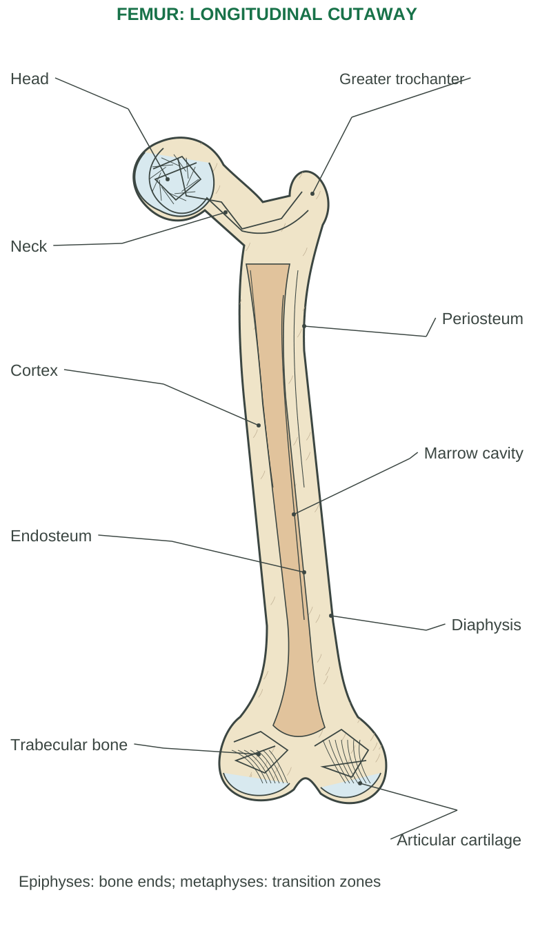
Diagram labels: Head; greater trochanter; neck; periosteum; cortex; marrow cavity; endosteum; diaphysis; trabecular bone; articular cartilage. Epiphyses: bone ends; metaphyses: transition zones.
Regions of a Long Bone
The diaphysis is the shaft. Epiphyses are the ends. Metaphyses connect shaft and ends and contain the growth plate in a growing bone. After growth plate closure, an epiphyseal line may mark the former region.
Articular cartilage covers participating joint surfaces and limits friction. The periosteum covers external non-articular surfaces and contains supportive and growth/repair-related elements. The endosteum lines internal bone surfaces.
Marrow and Blood Supply
A medullary cavity occupies much of the shaft of many long bones. Marrow distribution changes with age and differs by location. Nutrient vessels and additional regional vessels support bone cells. Do not draw a long-bone shaft as entirely solid without a marrow space.
| Cell | Main role |
|---|---|
| Osteogenic precursor | Gives rise to osteoblast lineage |
| Osteoblast | Produces osteoid and supports mineralization |
| Osteocyte | Mature embedded cell; senses mechanical environment and maintains/signals within tissue |
| Osteoclast | Multinucleated cell that resorbs bone |
Matrix
Type I collagen contributes tensile properties. Mineral, mainly hydroxyapatite-associated calcium phosphate, contributes rigidity and resistance to compression. Bone is strong because these components cooperate.
2.14 Microscopic Bone, Growth and Remodelling
Compact Bone and Osteons
Many regions of compact bone contain osteons: concentric lamellae around a central canal. Osteocytes occupy lacunae; canaliculi permit communication through cell processes. Perforating canals connect vessel routes across the bone.
Spongy Bone
Trabeculae align with mechanical demands. Their marrow spaces reduce weight while supporting a framework. Trabecular bone generally does not contain the same complete osteon arrangement as typical compact cortical bone.
Formation and Growth
Intramembranous ossification forms bone directly from mesenchymal tissue. Endochondral ossification replaces a cartilage model. Longitudinal growth of many long bones depends on activity at epiphyseal plates; width increases through appositional growth.
Remodelling
Resorption by osteoclasts and formation by osteoblasts continually renew bone. Mechanical loading and hormonal/metabolic conditions affect this balance. Repair of microdamage and mineral regulation occur alongside maintenance of strength.
Fracture Repair: Basic Sequence
Haematoma and inflammation → soft callus → bony callus → remodelling
These phases overlap. Proper stability and perfusion support repair; the exact course depends on fracture and host conditions.
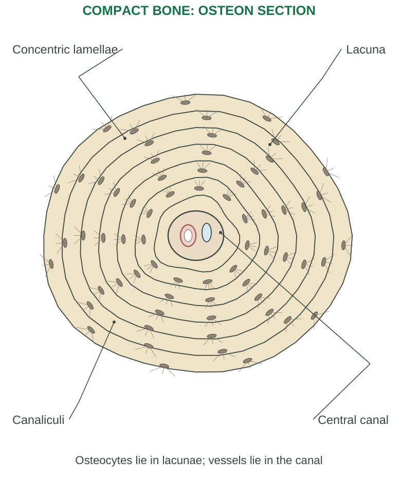
Diagram labels: Concentric lamellae; lacuna; canaliculi; central canal. Osteocytes lie in lacunae; vessels lie in the canal.
Cell-name memory: Blast builds; clast clears; cyte resides and communicates. This is a recall aid, not a substitute for understanding how the cells interact.
2.15 Axial Bones: Salient Features and Functions
Skull
The usual adult skull has 22 bones: 8 cranial and 14 facial. Cranial bones are frontal, paired parietal, paired temporal, occipital, sphenoid and ethmoid.
Facial bones include paired maxillae, zygomatic, nasal, lacrimal, palatine and inferior nasal conchae, plus vomer and mandible.
Most skull bones are joined by sutures. The mandible is mobile at the temporomandibular joints. The skull protects the brain and supports facial structures, teeth and sensory-organ spaces.
Hyoid and Auditory Ossicles
The hyoid supports tongue-related and swallowing structures and does not directly articulate with another bone. Each middle ear contains malleus, incus and stapes; these transmit sound-related mechanical vibration.
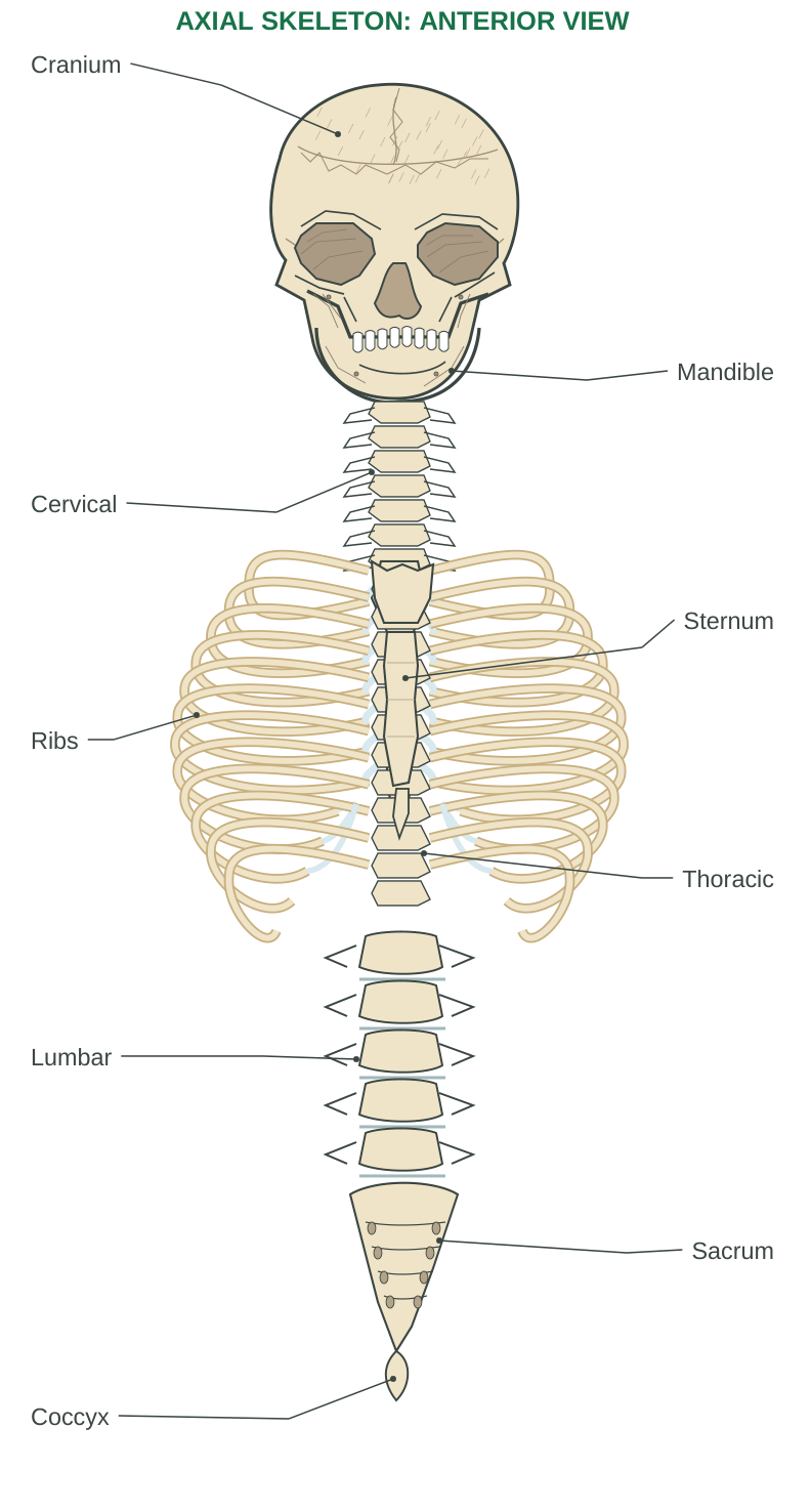
Diagram labels: Cranium; mandible; cervical; sternum; ribs; thoracic; lumbar; sacrum; coccyx.
Vertebral Column
| Region | Typical vertebrae | Important feature |
|---|---|---|
| Cervical | 7 | Neck support; atlas and axis support head movement |
| Thoracic | 12 | Articulate with ribs |
| Lumbar | 5 | Large bodies bear substantial load |
| Sacral | 5 fused | Transfer load toward the pelvis |
| Coccygeal | Usually 4 fused | Small terminal segment; number varies |
The adult column usually has 26 separate bones after sacral/coccygeal fusion. It supports body weight, protects the spinal cord and permits controlled movement. Intervertebral discs between most vertebral bodies contain an annulus fibrosus around a nucleus pulposus.
Thoracic Cage
Twelve pairs of ribs and the sternum form the anterior/lateral cage with thoracic vertebrae posteriorly.
- True ribs 1–7: Attach directly through their costal cartilages.
- False ribs 8–12: Lack direct attachment.
- Floating ribs 11–12: Have no anterior sternal attachment.
The sternum has manubrium, body and xiphoid process.
2.16 Appendicular Bones: Girdles and Limbs
Pectoral Girdle and Upper Limb
Each pectoral girdle has a clavicle and scapula. The clavicle acts as a strut; the scapula provides broad muscle attachment and the glenoid cavity for the humeral head. The arrangement permits high shoulder mobility.
| Region | Bones and salient relationship |
|---|---|
| Arm | Humerus; proximal head articulates with scapula |
| Forearm | Radius laterally on the thumb side; ulna medially in anatomical position |
| Wrist | 8 carpals per side |
| Palm | 5 metacarpals per side |
| Digits | 14 phalanges per hand: thumb 2, each other digit 3 |
Pronation/supination changes the relationship of radius and ulna. The hand supports fine manipulation through varied joints and muscular control.
Pelvic Girdle and Lower Limb
Each hip bone consists of fused ilium, ischium and pubis. The acetabulum receives the femoral head. The two hip bones form the pelvic girdle; with sacrum and coccyx they contribute to the bony pelvis. The pelvis supports/transmits weight and protects pelvic contents.
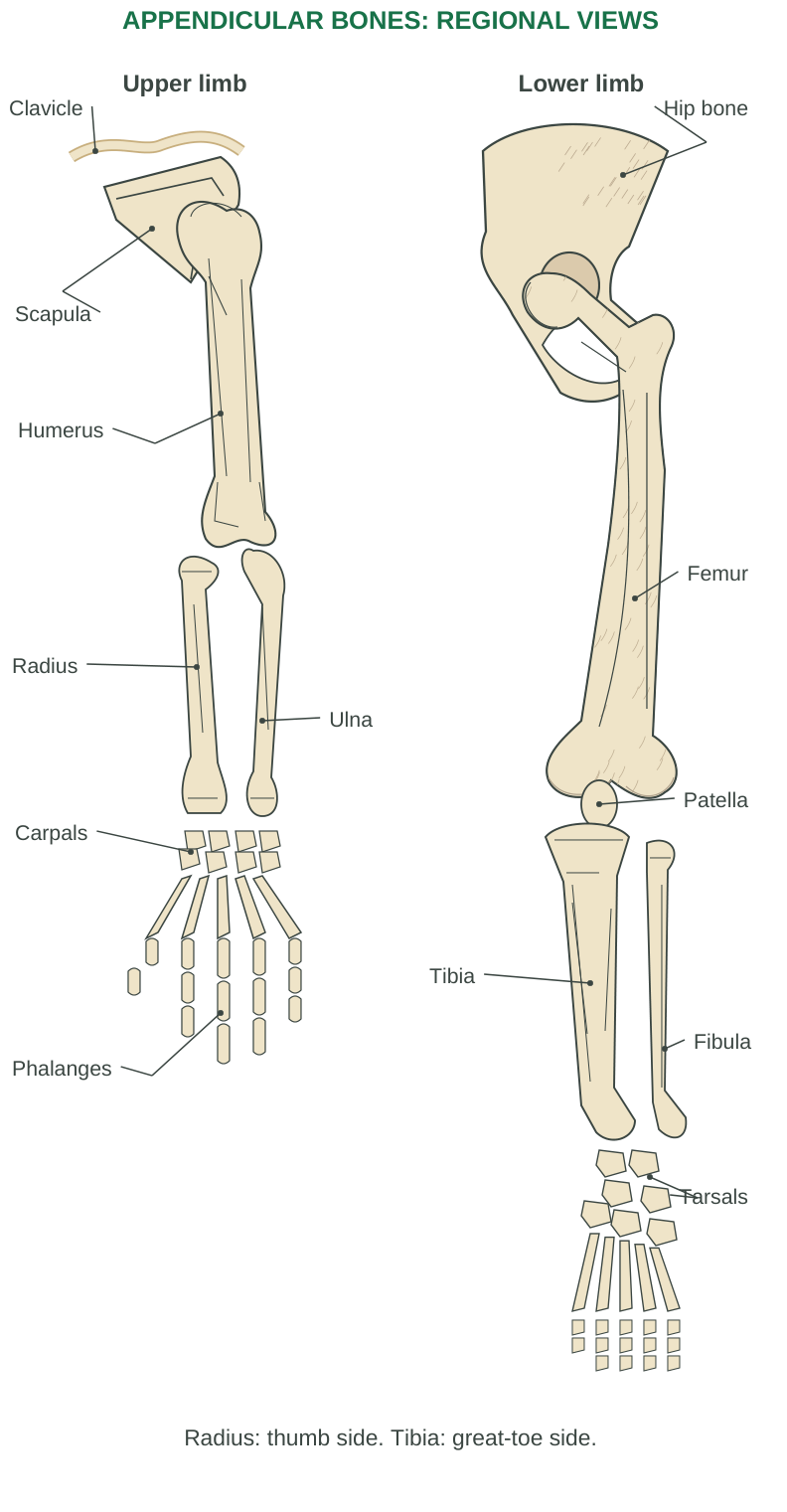
Diagram labels: Upper limb; lower limb; clavicle; scapula; humerus; radius; ulna; carpals; phalanges; hip bone; femur; patella; tibia; fibula; tarsals. Radius: thumb side. Tibia: great-toe side.
| Region | Bones and salient relationship |
|---|---|
| Thigh | Femur, strong weight-bearing long bone |
| Knee region | Patella, a sesamoid bone in the extensor mechanism |
| Leg | Tibia is the major medial weight-bearing bone; fibula is lateral |
| Ankle/hindfoot | 7 tarsals per side; talus receives load, calcaneus forms heel |
| Foot | 5 metatarsals and 14 phalanges per side |
Foot arches distribute load and aid locomotion. The lower limb emphasizes stability and weight transfer, while the upper limb generally emphasizes mobility and manipulation.
Terminology check: Anatomical arm means shoulder to elbow; leg means knee to ankle. These terms do not mean the entire upper or lower limb.
2.17 Skeletal Muscle: Organization and Coverings
A skeletal muscle consists of bundles of muscle fibres, connective tissue, vessels and nerves. A muscle fibre is an elongated multinucleated cell. It is not the same as a whole muscle or a fascicle.
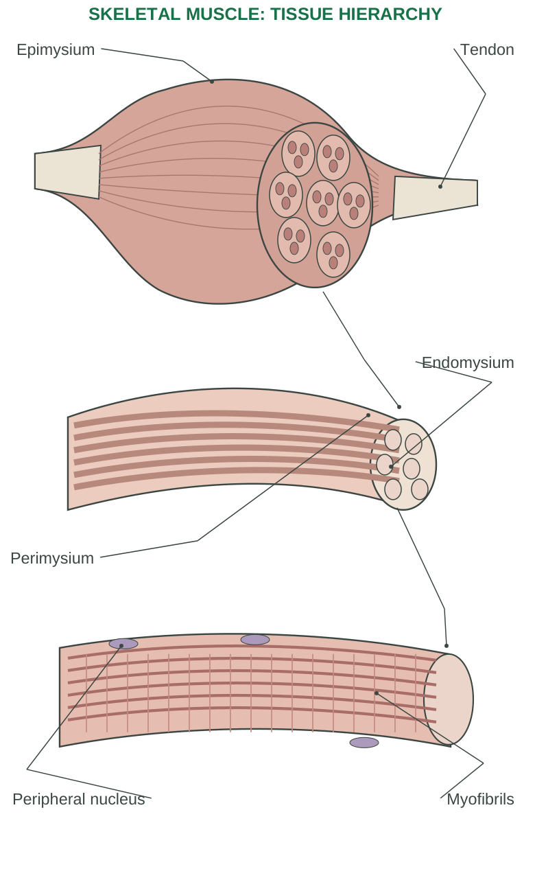
Diagram labels: Epimysium; tendon; perimysium; endomysium; peripheral nucleus; myofibrils.
| Level | Definition or covering |
|---|---|
| Whole muscle | Many fascicles; surrounded by epimysium |
| Fascicle | Bundle of muscle fibres; surrounded by perimysium |
| Muscle fibre | One cell; supported by endomysium |
| Myofibril | Contractile cylinder within a fibre |
| Sarcomere | Repeating unit between two Z discs |
| Myofilaments | Thin actin-associated and thick myosin filaments |
Force Transmission
Connective coverings integrate force from fibres and transmit it toward tendons or aponeuroses. A tendon commonly attaches muscle to bone, permitting force to act across a joint. The attachment is mechanically continuous with the supporting tissue; it is not merely a loose cord touching the bone.
Cellular Structures
The sarcolemma is the fibre membrane; sarcoplasm is the intracellular material. Many nuclei lie near the periphery. T-tubules carry surface excitation inward. The sarcoplasmic reticulum stores and regulates calcium required for contraction.
Structural Advantage
Long fibres and organized myofibrils generate directed tension. Vessels deliver substrates and oxygen, while neural input coordinates activation. A muscle may generate tension while shortening, remaining the same length or lengthening under load.
2.18 Sarcomere and Contractile Proteins
A sarcomere extends from one Z disc to the next. Its patterned thick and thin filaments produce skeletal-muscle striations.
| Structure | Main identity |
|---|---|
| Z disc | Sarcomere boundary; anchors thin filaments |
| Thin filament | Actin with regulatory troponin and tropomyosin |
| Thick filament | Myosin molecules with projecting heads |
| A band | Length of thick filaments, including overlap regions |
| I band | Thin-filament region without thick filaments |
| H zone | Central thick-filament region without thin overlap |
| M line | Central organizing region of thick filaments |
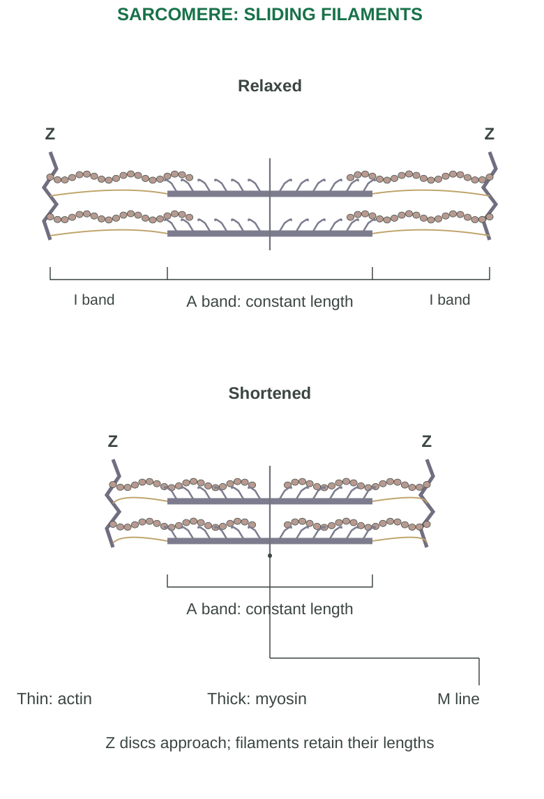
Diagram labels: Relaxed; shortened; Z discs; A band: constant length; I band; thin: actin; thick: myosin; M line. Z discs approach; filaments retain their lengths.
Regulatory Proteins
Tropomyosin restricts access to myosin-binding sites on actin in the resting state. Calcium binds the appropriate troponin subunit, changing the position of the regulatory complex and permitting productive actin–myosin interaction. Titin contributes organization and elastic properties.
Sliding Filament Principle
Thin filaments slide relative to thick filaments. Filament lengths do not shorten. Z discs approach; the I band and H zone narrow, while A-band length remains essentially unchanged. Under some conditions the H zone can disappear.
Learn by Tracing Boundaries
Draw two Z discs, a central M line and a symmetric thick-filament region. Add thin filaments from both ends. Mark A band across the entire thick-filament length, rather than labelling only the region of thick/thin overlap as A band.
Exam trap: A myofibril is made of repeating sarcomeres. The whole muscle fibre contains many myofibrils, organelles and membranes; it is not one giant sarcomere.
2.19 Neuromuscular Junction (NMJ)
The NMJ is the specialized synapse between a somatic motor neuron terminal and a skeletal muscle fibre. The muscle membrane beneath the terminal forms a motor end plate with junctional folds.
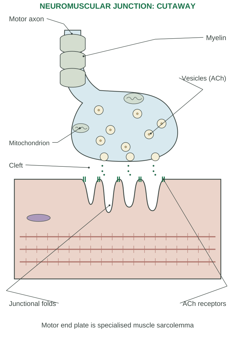
Diagram labels: Motor axon; myelin; vesicles (ACh); mitochondrion; cleft; junctional folds; ACh receptors. Motor end plate is specialised muscle sarcolemma.
Main Components
The presynaptic terminal contains acetylcholine (ACh) vesicles and mitochondria. A synaptic cleft separates neuron and muscle. The postsynaptic membrane contains muscle-type nicotinic ACh receptors. Acetylcholinesterase in the cleft region terminates the transmitter’s action.
Stepwise Transmission
- Step 1: A neuronal action potential reaches the terminal.
- Step 2: Voltage-gated calcium channels open; calcium enters the terminal.
- Step 3: Vesicles fuse and release ACh by exocytosis.
- Step 4: ACh binds nicotinic receptors; ligand-gated cation channels open.
- Step 5: The end-plate potential depolarizes nearby membrane enough to activate voltage-gated sodium channels and generate a muscle action potential.
- Step 6: ACh is hydrolysed; choline can be taken up for reuse.
Potential Distinction
An end-plate potential is local and graded. A muscle action potential is propagated. ACh receptors are not themselves identical to the voltage-gated sodium channels that propagate the action potential.
Motor Unit
One motor neuron plus all muscle fibres it supplies is a motor unit. Small units permit fine control; larger units can recruit more fibres together. Increasing motor-unit recruitment increases force in a whole muscle.
2.20 Excitation–Contraction Coupling
Excitation–contraction coupling connects a muscle action potential to calcium-regulated force generation. It is the bridge between electrical signalling and mechanical activity.
Skeletal-Muscle Sequence
- Step 1: The action potential spreads along sarcolemma and enters T-tubules.
- Step 2: T-tubule voltage sensing activates nearby SR calcium-release channels through specialized coupling.
- Step 3: Calcium moves from SR into sarcoplasm.
- Step 4: Calcium binds troponin, allowing tropomyosin to move from blocking positions.
- Step 5: Myosin interacts cyclically with actin while ATP is available.
Triad
A skeletal-muscle triad consists of one T-tubule and two adjacent terminal cisternae of SR. The T-tubule is continuous with the surface membrane. The SR is a separate intracellular calcium-storage system; the two are closely related but are not one continuous hollow tube.
Relaxation
When excitation stops, calcium is pumped back into SR, mainly by SERCA ATPases. Cytosolic calcium falls, troponin loses activating calcium and tropomyosin again limits binding. Elastic and external forces help restore length when the mechanical situation allows.
Calcium Versus ATP
Calcium permits activation; ATP supplies energy for the cross-bridge cycle and powers calcium reuptake and membrane-gradient maintenance. Calcium is not a substitute fuel for ATP.
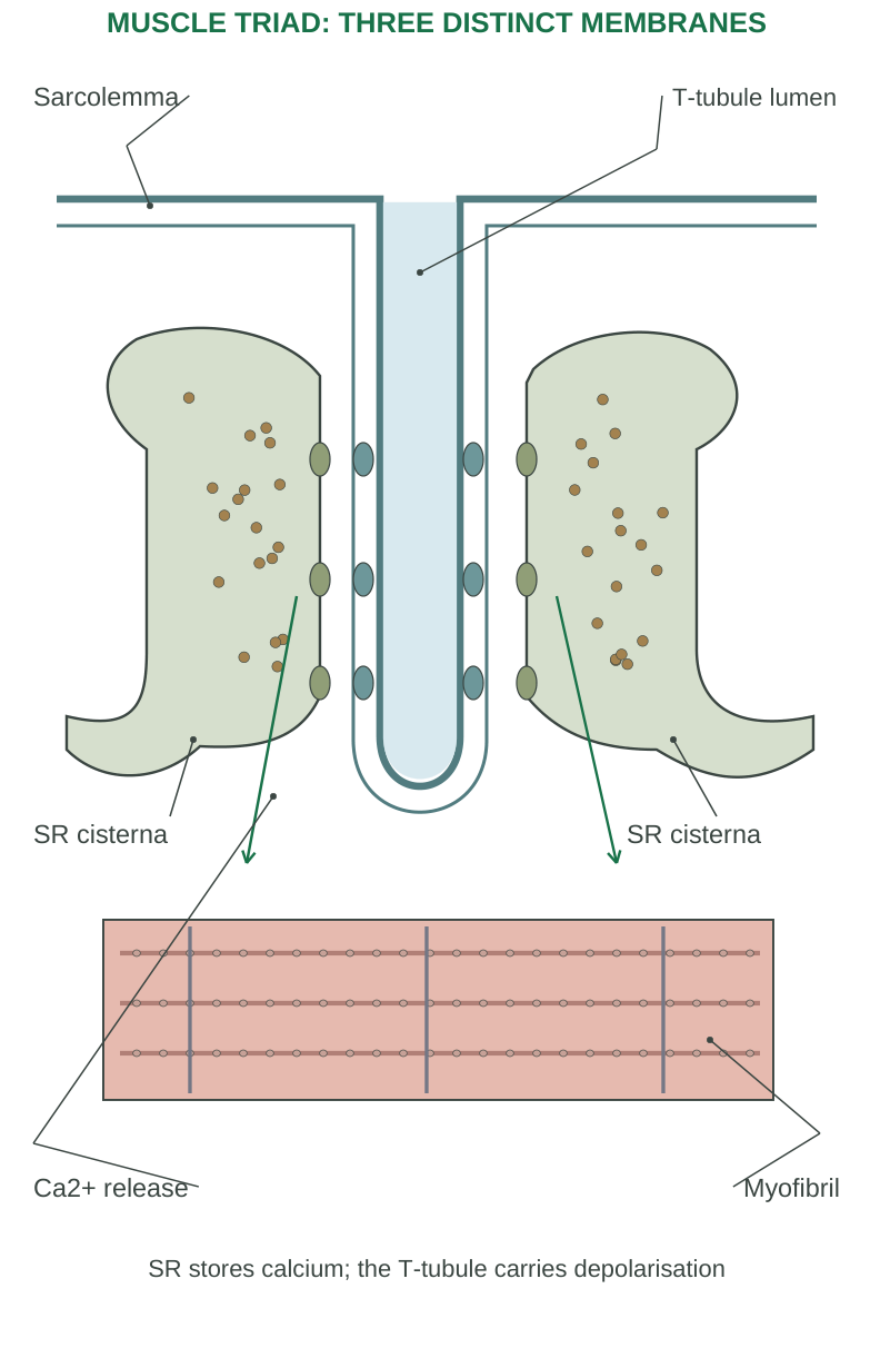
Diagram labels: Sarcolemma; T-tubule lumen; two SR cisternae; Ca2+ release; myofibril. SR stores calcium; the T-tubule carries depolarisation.
Skeletal-muscle qualification: Do not transfer every cardiac or smooth-muscle calcium mechanism directly into this answer. Skeletal muscle has its own specialized T-tubule–SR coupling.
2.21 Cross-Bridge Cycle and ATP Functions
Four Key Events
- Attachment: An energized myosin head binds an exposed actin site.
- Power stroke: Release of phosphate and subsequent changes move the head, drawing actin relative to myosin; ADP is released.
- Detachment: Fresh ATP binding lowers myosin’s affinity for actin.
- Re-cocking: ATP hydrolysis energizes/repositions the head for another cycle.
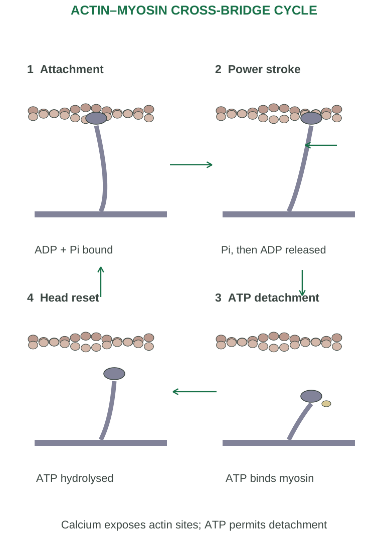
Diagram labels: Attachment: ADP + Pi bound. Power stroke: Pi, then ADP released. ATP detachment: ATP binds myosin. Head reset: ATP hydrolysed. Calcium exposes actin sites; ATP permits detachment.
Conditions for Repeated Cycling
Accessible actin sites and ATP are needed. Individual myosin heads are not all synchronized, allowing sustained tension through many overlapping interactions. When calcium is removed and sites become less available, productive cycling declines.
| ATP-related event | Why it matters |
|---|---|
| ATP binds myosin | Permits detachment from actin |
| ATP is hydrolysed | Primes the head for another cycle |
| SR calcium pumping | Supports relaxation |
| Ion pumping | Maintains gradients required for excitation |
Rigor Concept
Without ATP, myosin cannot normally detach and calcium regulation fails. This helps explain rigor after death. It does not mean that the absence of ATP is the normal physiological signal to contract.
Mechanical Distinction
A cross-bridge cycle is a molecular event; a twitch is a mechanical response of a fibre/muscle to one stimulus; sustained whole-muscle force depends on many molecular cycles, activation timing and motor-unit recruitment.
Memory line: Bind → pull → detach → reset. ATP binding is particularly associated with detachment; do not incorrectly state that binding ATP directly creates the power stroke.
2.22 Twitch, Summation and Forms of Contraction
A Single Twitch
The latent period reflects events before detectable force. The contraction phase raises tension; the relaxation phase lowers it as activation subsides. The electrical action potential is much briefer than the mechanical twitch.
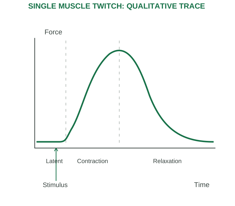
Diagram labels: Force; time; stimulus; latent; contraction; relaxation.
Summation and Tetanus
If a new stimulus arrives before complete relaxation, force responses can summate. Increasing stimulation frequency may produce incomplete then fused tetanus. Physiological muscle tetanus is sustained force, not the infectious disease tetanus.
Recruitment and Muscle Tone
Whole-muscle force increases by activating more motor units and changing firing patterns. Muscle tone is a background level of activity contributing to posture and readiness; it is not a state of maximum contraction.
| Form | Basic mechanical description |
|---|---|
| Isometric | Tension changes with little/no overall muscle-length change |
| Concentric | Muscle shortens while generating force |
| Eccentric | Activated muscle lengthens under load |
| Isotonic idealization | Length changes while load/tension is approximately constant |
Length and Load
Starting sarcomere length influences productive filament overlap. Too little or excessive overlap reduces potential active force. Load affects shortening, and muscle activation may generate tension without movement if the external load is not overcome.
Energy Supply and Fatigue
ATP is replenished through phosphocreatine transfer, glycolysis and oxidative metabolism. Fatigue is multifactorial: altered excitation, calcium handling, metabolic conditions and neural factors can contribute. It is not correctly explained only as “all ATP disappears.”
2.23 Structural and Functional Classification
A joint is an articulation between skeletal elements. Structural classification concerns connecting material and cavity; functional classification concerns permitted movement.
| Structural group | Arrangement | Example |
|---|---|---|
| Fibrous | Fibrous connective tissue, no synovial cavity | Skull suture; distal tibiofibular syndesmosis; tooth gomphosis |
| Cartilaginous | Cartilage connects elements, no synovial cavity | Growth-plate synchondrosis; pubic symphysis |
| Synovial | Articular surfaces separated by a fluid-containing joint cavity | Knee, hip, shoulder |
Fibrous Joints
Sutures join most skull bones. Syndesmoses connect bones through ligaments or membranes and can allow limited movement. Gomphoses secure teeth in sockets. These three arrangements should not all be described as completely immovable in every circumstance.
Cartilaginous Joints
Synchondroses join bones through hyaline cartilage; some are temporary growth-related structures. Symphyses involve fibrocartilage and provide strength with limited movement, as at the pubic symphysis and between vertebral bodies through discs.
| Functional term | Meaning | Common illustration |
|---|---|---|
| Synarthrosis | Essentially immovable | Many sutures |
| Amphiarthrosis | Slightly movable | Pubic symphysis |
| Diarthrosis | Freely movable | Synovial joint |
Two Classifications, Different Questions
Do not equate every fibrous joint with absolute immobility. Structure and function correlate but are not identical naming systems. Synovial joints are functionally diarthroses, with their precise motion limited by shape and supporting structures.
2.24 Synovial Joint Structure and Types
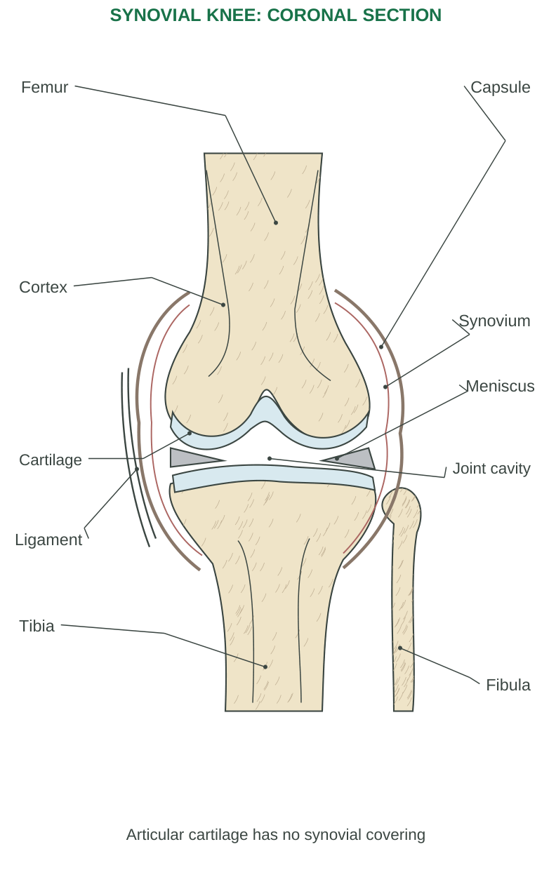
Diagram labels: Femur; capsule; cortex; synovium; cartilage; joint cavity; tibia; fibula; ligament; meniscus. Articular cartilage has no synovial covering.
Essential Structures
Articular cartilage, usually hyaline, covers contacting bone ends. A joint cavity contains synovial fluid. The capsule has an outer fibrous layer and an inner synovial membrane lining appropriate internal non-articular surfaces. Synovial fluid lubricates and helps nourish avascular cartilage. Ligaments stabilize relationships. Some joints contain discs/menisci or bursae; these are accessory structures, not mandatory parts of every synovial joint.
| Type | Main movement | Example |
|---|---|---|
| Plane | Gliding | Many intercarpal joints |
| Hinge | Mainly flexion/extension | Humeroulnar elbow component |
| Pivot | Rotation about an axis | Proximal radioulnar joint |
| Condyloid | Biaxial motion | Radiocarpal wrist |
| Saddle | Biaxial motion with reciprocal surfaces | Thumb carpometacarpal joint |
| Ball-and-socket | Multiaxial motion | Hip and shoulder |
Movement Terms
Flexion generally reduces the angle; extension increases it. Abduction moves away from the midline/reference axis; adduction returns toward it. Rotation turns around an axis. Circumduction combines movements into a cone-like path. Pronation/supination involve the forearm; dorsiflexion/plantarflexion refer to the ankle region.
Stability–Mobility Relationship
Joint shape, capsule/ligaments, muscles and neural control interact. Greater mobility may require more active stabilization. A bone’s articular shape alone does not explain every movement limit.
Trace the sketch: Point to cartilage, synovial fluid space, synovial lining and outer capsule. Do not label the cartilage itself as the synovial membrane.
2.25 Rheumatoid Arthritis (RA)
RA is a chronic systemic autoimmune inflammatory disease with prominent synovial-joint involvement. It is different from simple age-related cartilage wear.
Pathogenesis
Susceptibility and environmental influences contribute to loss of immune tolerance. Autoimmune responses, immune cells and inflammatory mediators such as TNF and IL-6 sustain synovitis. The inflamed lining becomes hyperplastic and vascular, and destructive pannus can extend over cartilage and erode bone.
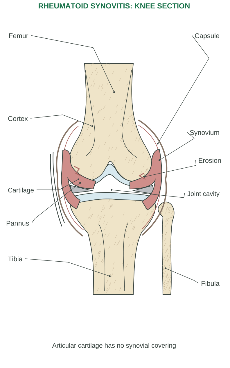
Diagram labels: Femur; capsule; cortex; synovium; cartilage; joint cavity; tibia; fibula; pannus; erosion. Articular cartilage has no synovial covering.
Tissue-to-Function Sequence
Persistent synovitis → swelling/pain → cartilage and marginal bone injury → altered joint mechanics → deformity and loss of function
Tendons, ligaments and surrounding tissues can also be affected. The degree and pattern vary across individuals.
Typical Findings
Small hand, wrist and foot joints are commonly affected, often symmetrically. Prolonged morning stiffness, swelling and fatigue are useful features. Systemic manifestations may involve organs outside the joints; nodules occur in some patients.
Laboratory Interpretation
Rheumatoid factor and anti-citrullinated protein antibodies can support classification and assessment, but a positive result is not an automatic diagnosis and negative tests do not completely exclude disease. Interpret clinical, laboratory and imaging evidence together.
Disease Modification Concept
Suppressing pain is different from limiting immune-mediated structural injury. In examinations, connect treatment principles to the mechanism without substituting a drug list for the requested pathophysiology.
Key image distinction: The abnormal lining/pannus is central. Do not draw RA merely as two rough bone ends with the cartilage unchanged.
2.26 Osteoporosis: Loss of Bone Strength
Osteoporosis is reduced bone strength associated with reduced bone mass and deterioration of microarchitecture, increasing fragility-fracture risk. The remaining bone is generally normally mineralized; this distinguishes it from defective mineralization in osteomalacia.
Mechanism
Bone resorption can exceed formation over time. Reduced peak bone mass or accelerated later loss may contribute. Estrogen decline, aging, reduced loading, glucocorticoid exposure and other secondary causes can alter the balance. Trabeculae become thinner or disconnected and cortical structure can weaken.
Consequences
Vertebral, hip and wrist fractures are important examples. Vertebral compression can cause loss of height, deformity and pain. The condition may remain clinically silent until a fracture occurs; absence of bone pain does not prove normal strength.
Assessment Concept
Bone mineral density is one component of fracture-risk assessment. A T-score compares a result with a young-adult reference population. Its interpretation depends on age and clinical context; it is not a universal classification rule for every child or young adult.
Protective Principles
Appropriate loading/activity, adequate nutritional support, attention to secondary causes and reduction of falls support bone health. Calcium alone does not automatically correct every mechanism of osteoporosis.
Comparison
- Osteoporosis: Insufficient amount/architecture of bone with generally normal mineralization.
- Osteomalacia: Defective mineralization.
- Arthritis: Joint disease.
These are not interchangeable terms.
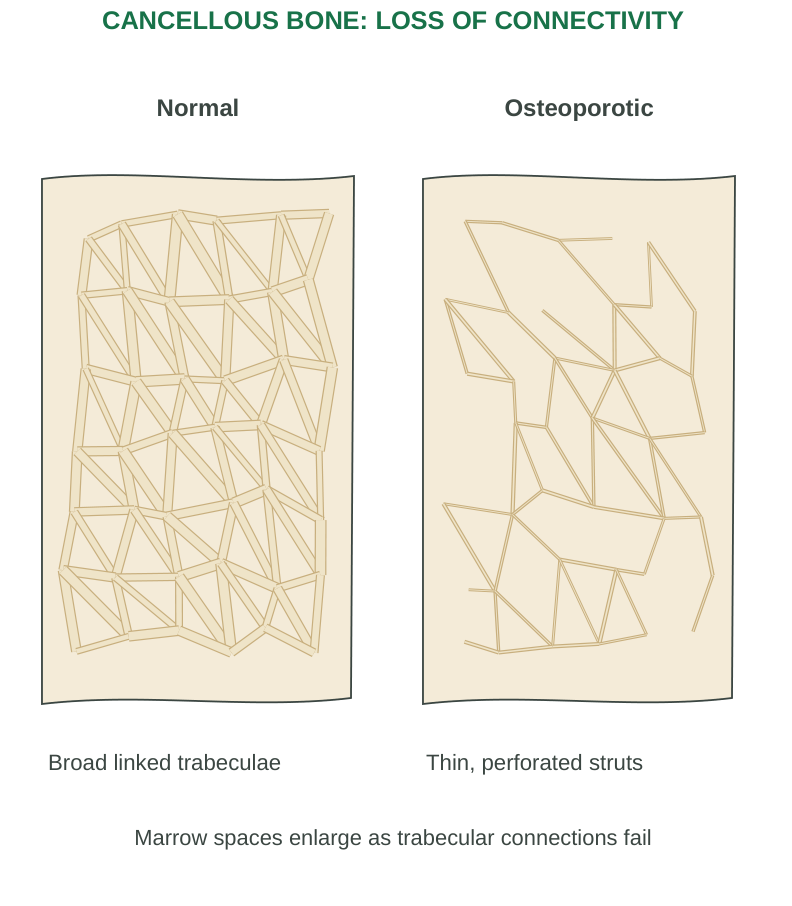
Diagram labels: Normal: broad linked trabeculae. Osteoporotic: thin, perforated struts. Marrow spaces enlarge as trabecular connections fail.
2.27 Gout: Urate Crystals and Inflammation
Gout is an inflammatory disorder caused by deposition of monosodium urate crystals. Persistent elevated urate increases the likelihood of deposition, but hyperuricaemia alone does not mean that every person has clinical gout.
Pathogenesis
Urate arises from purine metabolism. Reduced renal/intestinal elimination or increased production can increase the body urate burden. When local conditions permit crystal formation, innate immune responses—including inflammasome-related IL-1 signalling—recruit inflammatory cells and produce a painful flare.
Manifestations and Progression
A flare commonly causes sudden severe pain, swelling and warmth in a joint; the first metatarsophalangeal joint is a classic site, but many other sites can be affected. Recurrent crystal burden may produce tophi, chronic joint damage and associated urinary stone problems in some people.
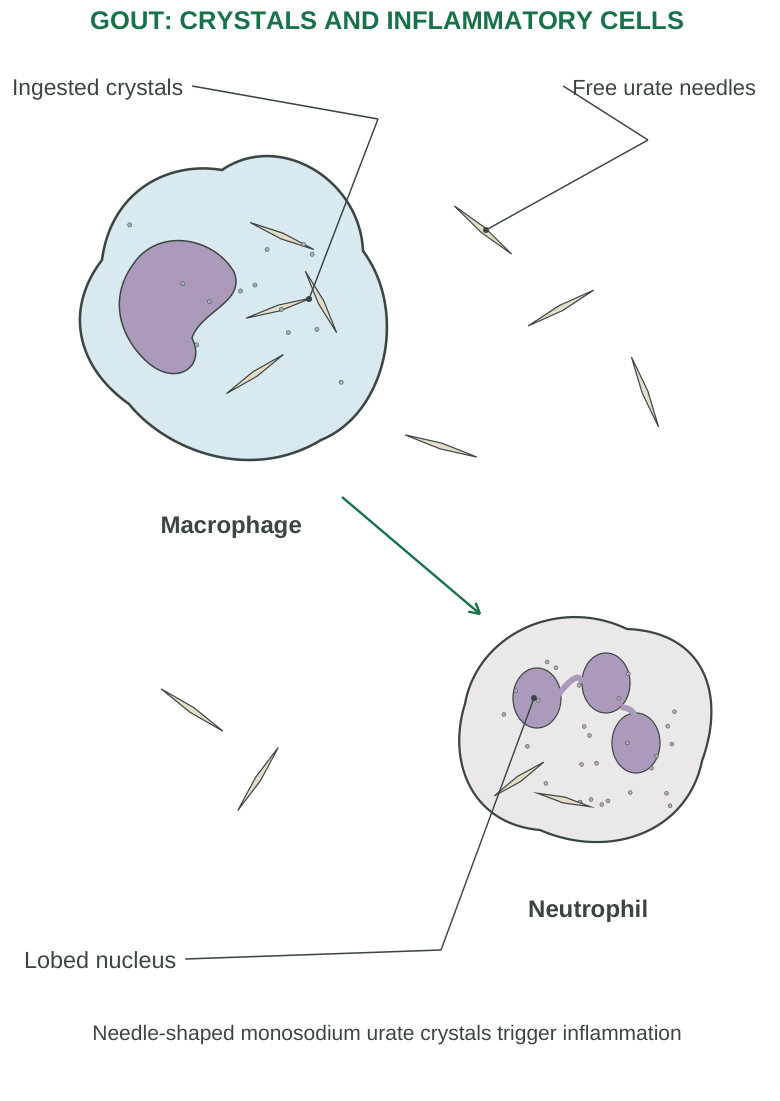
Diagram labels: Macrophage; neutrophil; ingested crystals; free urate needles; lobed nucleus. Needle-shaped monosodium urate crystals trigger inflammation.
Crystal Identification
Monosodium urate crystals are typically needle-shaped and negatively birefringent under compensated polarized microscopy. These features distinguish them from calcium pyrophosphate crystals. Serum urate alone is insufficient to establish every acute-joint diagnosis and may not be high during a flare.
Mechanism-Based Distinction
Acute inflammation and the long-term urate burden are related but separate targets. Relief of a flare does not by itself prove that deposited crystals have been eliminated.
Three disease mechanisms: RA: autoimmune synovitis. Osteoporosis: reduced bone strength. Gout: urate-crystal inflammation. Explain the initiating tissue process before listing symptoms.
2.28 Worked Cases and Rapid Revision
Case 1: A Wound Extends into Dermis
A deeper skin cut may bleed because the dermis contains vessels. Loss of connective tissue increases the need for matrix repair and scar formation. Epidermis alone is avascular.
Case 2: Muscle Fails to Relax Normally
Restoration of low sarcoplasmic calcium requires ATP-powered SR pumping. ATP also permits myosin detachment. Therefore, lack of ATP affects both molecular cycling and relaxation; it is not merely a loss of “pulling power.”
Case 3: Identify the Band Change
A sarcomere shortens from 2.5 micrometres to 2.0 micrometres.
Percentage shortening = (2.5 − 2.0)/2.5 × 100 = 20%.
The A band remains approximately constant; I band/H zone narrow. This calculation describes length change, not force.
Case 4: Count Bones in One Hand
8 carpals + 5 metacarpals + 14 phalanges = 27 bones.
The thumb contributes 2 phalanges and the other four digits contribute 12. Do not confuse one hand’s count with the entire upper limb.
Case 5: Match Mechanism and Disorder
- Scaly plaque with immune-related epidermal changes: psoriasis mechanism.
- Barrier disruption with itch–scratch amplification: dermatitis mechanism.
- Sensory loss with peripheral nerve involvement: consider leprosy-related injury.
Clinical diagnosis still requires appropriate evidence.
Compare Joint Diseases
| Feature | RA | Osteoporosis | Gout |
|---|---|---|---|
| Main tissue process | Autoimmune synovitis | Weak bone architecture | Urate-crystal inflammation |
| Structural result | Cartilage/bone erosion | Fragility and fractures | Deposits and possible erosions |
| Typical teaching clue | Symmetric inflammatory joints | Low-trauma fracture | Acute painful swollen joint |
One-Minute Recall
- Skin: Barrier and sensation.
- Bone: Support, minerals and marrow.
- Muscle: ATP-dependent force.
- Joint: Articulation and controlled motion.
Always add a structure and mechanism to the one-word function.
2.29 Examination Practice
Long-Answer Questions
- 1. Draw a labelled skin section and explain layer-specific functions.
- 2. Explain psoriasis, dermatitis and leprosy through their mechanisms.
- 3. Describe the four overlapping phases of wound healing and factors that modify repair.
- 4. Classify bones and describe axial and appendicular divisions with examples and functions.
- 5. Explain skeletal-muscle organization and the sliding filament mechanism.
- 6. Draw and explain the NMJ; trace excitation–contraction coupling and relaxation.
- 7. Classify joints structurally and functionally; describe a synovial joint.
- 8. Compare the pathophysiology of RA, osteoporosis and gout.
Short-Answer Practice
- Epidermal strata and cells; thick versus thin skin.
- Eccrine versus sebaceous glands.
- Granulation tissue and scar maturation.
- Primary versus secondary intention.
- Bone cells, osteon and long-bone regions.
- Radius versus ulna; tibia versus fibula.
- Sarcomere bands and ATP functions.
- Motor unit and end-plate potential.
- Hinge, pivot, saddle and ball-and-socket joints.
- Pannus, tophus and fragility fracture.
Quick Test: Answers Below
- 1. Which skin layer lacks blood vessels?
- 2. Which bone cell resorbs matrix?
- 3. Which sarcomere band stays essentially the same length during shortening?
- 4. What binds troponin to permit activation?
- 5. What event permits myosin to detach from actin?
- 6. Which joint lining is prominently inflamed in RA?
- 7. Which crystal causes gout?
Answers:
- 1. Epidermis.
- 2. Osteoclast.
- 3. A band.
- 4. Calcium.
- 5. ATP binding.
- 6. Synovium.
- 7. Monosodium urate.
Worked Distinction
Question: Why can a small phalanx be a long bone, while a larger cranial bone is flat?
Answer: Shape/architecture determines classification. A phalanx has a shaft and ends with length exceeding width. A cranial vault bone forms a relatively thin protective plate.
Final Checklist
- Trace skin layers and label appendages clearly.
- Distinguish all three required skin disorders.
- Sketch wound changes across four phases.
- Recall axial/appendicular bones and their functions.
- Label long bone, osteon, muscle fibre and sarcomere.
- Explain NMJ → calcium release → cycling → relaxation.
- State band changes without shortening filaments.
- Compare structural and functional joint groups.
- Explain all three bone/joint diseases through mechanisms.
Unit II Complete
Practise one anatomical sketch, one process sketch and one mechanism-based answer each revision session.